The Visual Map Is Redrawn Before It Reaches the Cortex

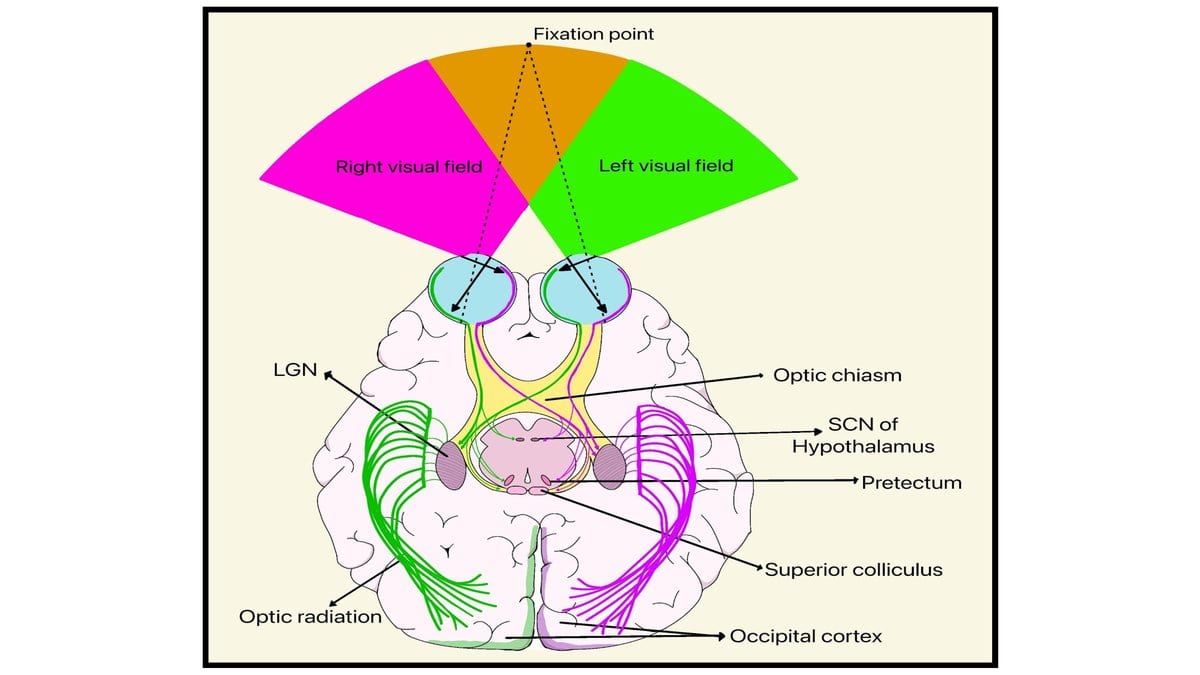
Keep your eyes on this word and notice, without moving them, what is happening at the outer limit of what you can see. A doorway to the left. Someone crossing the room at the far right. That band of vision is coarse, nearly colorless, and most of the time you barely attend to it at all.
For people who have been deaf since early childhood, it does more work. Tests going back to the 1980s have found that deaf adults are better than hearing adults at catching things out there, at the far edge. What a team at the Universities of York and Sheffield wanted to know was whether the brain's own layout has shifted to match, and how far down the visual pathway any shift reaches.
Their answer, published Sept. 4 in the Proceedings of the National Academy of Sciences, is that it reaches deeper than the cortex.
Alexandra Levine, Heidi Baseler and colleagues put 16 adults who had been profoundly deaf from early in life into a scanner, alongside 16 hearing adults matched to them for age. The paper refers to the group as "D/deaf," a convention covering both the audiological fact and the cultural identity.
What they measured takes advantage of one of the visual system's oldest design features: it keeps the geometry of the world. Neighboring parts of a scene land on neighboring patches of tissue. So a functional MRI scan, which follows blood flow as a stand-in for activity, can be read as a chart of which square of brain is looking after which part of the view. Sixteen against sixteen is a small sample for that kind of comparison, small enough that the size of any difference is better treated as unsettled than as measured.
In the deaf group, the far periphery had more of the map. The center had less. Neither the visual cortex nor the relay beneath it differed between the groups in overall size, so nothing had grown; the proportions had moved. The authors describe it as a redistribution of neural resources.
That relay is the part of the result that had not been reported before. Signals from the eye do not go straight to the cortex; they stop at the lateral geniculate nucleus, a way station in the thalamus, deep in the middle of the brain. Most of the search for this kind of reorganization has been conducted in the cortex, where the visual map is easy to reach and easy to read. The York and Sheffield scans found the same lopsided proportions one stop earlier, in tissue long treated as a relay rather than as a place where a lifetime of use leaves a mark.
The cortical half of the finding, by contrast, is a confirmation. An independent group reported a similar peripheral bias in the visual cortex of congenitally deaf adults in 2016 (populations of cells there answered to wider patches of the far edge), and the new paper cites that work. Saying so does not shrink the result. It is what makes the thalamic finding land: the cortex was already known to be redrawn, and the open question was how much further down the redrawing went.
What a map's proportions describe is how tissue is allotted, which is not the same as what a person sees. The peripheral advantage itself comes from earlier behavioral work, not from these scans, and the link between the two is an inference. A redistributed map is not a report on anyone's experience of vision, or of deafness. It says that a visual system, used a particular way through childhood, ends up shaped that way; the framing here is about how much a part of the system was used, not about the brain repairing something missing.
The work itself is not new to the public record. A version of this paper has been readable as a preprint since January 2020; what changed this month is that it completed peer review and appeared in PNAS, open access, where anyone can read it.
One thing the scans cannot settle is which way the influence runs. The thalamus feeds the cortex and the cortex sends signals back down, so a bias measured in the relay may have been built there or may be an echo of the map above it. Telling those apart needs a study designed for the question. This one moves where that study has to look: below the cortex, one stop closer to the eye.
Sources
- Proceedings of the National Academy of SciencesPeer-reviewed
Medical Disclaimer: This content is provided for general informational and educational purposes only. It is not medical advice and should not be used as a substitute for professional medical advice, diagnosis, or treatment. Always consult a qualified healthcare professional regarding any medical condition, concern, symptom, or treatment decision.