Skip to content
See the World Through ScienceA project of ALLATRA
Source: Peer-reviewedNature1 source

Mice Can Now Swim and Compete While Their Brain Cells Are Recorded
Science

Republish this story

Our work is licensed under Creative Commons BY-NC 4.0. You may republish this piece for free — with credit to ALLATRA Media and a link to the original, unedited beyond length trims, and not for commercial use.

Read the full license

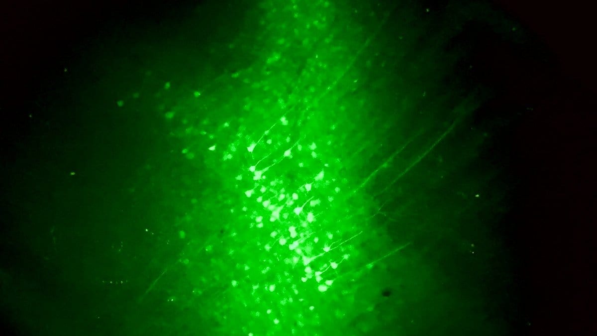
A dense cluster of nerve cells glowing bright green under a fluorescence microscope, their branching fibers spreading into darker surrounding tissue.
A fluorescent label makes individual cortical cells and their branches glow in brain tissue. Light like this is the signal a head-mounted microscope reads out while an animal moves (illustrative)."Cortical neurons" by Luisa Demarchi, Ilaria Bertocchi, Alessandra Oberto, via wikimedia, CC-BY-SA-4.0

A group at Tsinghua University has built a head-mounted microscope that weighs 1.0 gram and sends its data out wirelessly, and used it to watch brain cells in mice while the animals swam, explored mazes and competed with each other. The work was published Oct. 19, 2026, in Nature.

Microscopes small enough for a mouse to carry have had to be tethered by a cable and could cover only a narrow patch of brain, which limits what an animal is allowed to do while it is being recorded. The authors state that their device, which they call Airscope, makes it practical to record from several naturally behaving animals at once.

The team reports that Airscope resolves features down to 4 micrometers across a field of view 6 millimeters wide, at 12.5 frames a second, and that its wireless link carried calcium imaging, which tracks the flashes of light cells give off as they fire, from more than 8,500 neurons in a single animal. The paper gives that count as a 25-fold increase, and credits a lens design whose optical stack is 2.84 times shorter than in previous miniaturized microscopes.
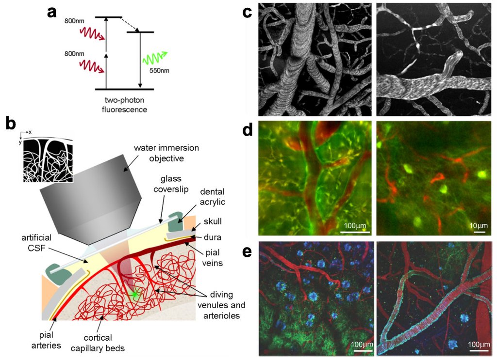
A multi-panel scientific figure: an energy diagram of two-photon fluorescence, a cutaway drawing of a microscope objective sitting over a glass window in the skull, and gray and color micrographs of blood vessels and cells in the cortex.
A glass window in the skull lets a benchtop microscope reach the cortex of a living animal, an arrangement that holds the head still (illustrative). "Two-photon microscopy of in vivo brain function" by Armen R Kherlopian, Ting Song, Qi Duan, Mathew A Neimark, Ming J Po, John K Gohagan and Andrew F Laine, via wikimedia, CC-BY-2.0

On what the instrument recorded, the authors report patterns of activity in the cortex, the brain's outer layer, that correlate with behavioral signs of social dominance, and population activity that decoded in advance which of two paired mice would win a contest. They also report activity tied to reward locations as mice explored a maze and, with two devices on one animal, a directional influence running from the spinal cord to the cortex during fear conditioning.

The first author is Yuanlong Zhang and the senior author is Qionghai Dai, both at Tsinghua University. The group also includes researchers at Fudan University, Princeton University, Northeastern University in Shenyang and the Chinese PLA General Hospital in Beijing.

Sources

By Olga SchmidtEditor-in-Chief, Writer

Spot an error?

Spot an error?

Report an error

Spotted a mistake on this page? Tell us what's wrong and our editors will take a look.

What kind of problem?

Only if you'd like us to be able to follow up. We won't use it for anything else.

We correct mistakes openly. Select any text to flag it. Fixes are logged under our Corrections Policy.

Report an error

Reporting on

Mice Can Now Swim and Compete While Their Brain Cells Are Recorded

What kind of problem?

Only if you'd like us to be able to follow up. We won't use it for anything else.

We read every report. Corrections are logged publicly.