In a Fish Larva's Eye, Worn Light Sensors Come Away in Small Pieces

The light-catching tip of a cone cell, its outer segment, is a dense stack of membrane, and it wears out from the far end. New material goes in at the base, the oldest material ends up at the top, and the top sits against a sheet of support cells at the back of the eye whose duties include carrying the worn material off. This has to work every day, in every cone, for as long as an animal can see. How it works inside a living eye has been much harder to establish than how much it matters.
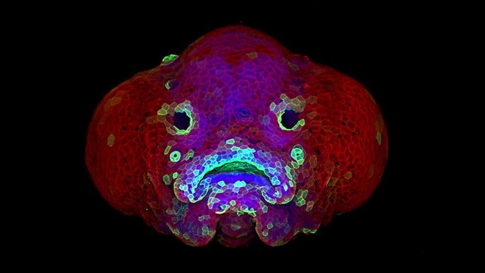
A preprint posted Oct. 25, 2026, reports watching it happen. Noora Emilia Nevala, Soile Nymark and their colleagues at Tampere University in Finland, working with two co-authors at Washington University School of Medicine in St. Louis, used live imaging of zebrafish larvae. They tagged the membranes of particular cone types and of the support layer separately, so the boundary between the two could be followed. The work has not been through peer review.
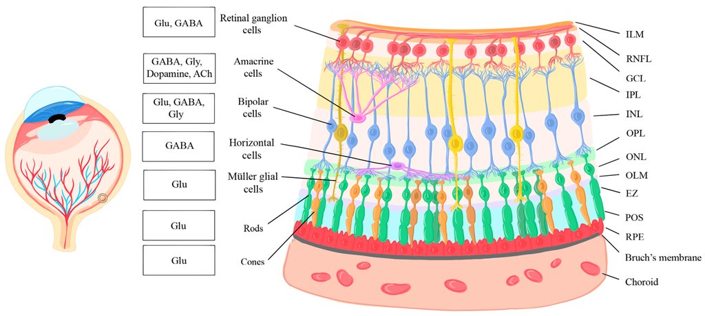
The sheet of cells behind the retina is the retinal pigment epithelium, a single layer packed with dark pigment. Its housekeeping job is long established: it takes in the oldest material from the tips of rods and cones and breaks it down. Several of the same authors reported in The FASEB Journal on July 24, 2025, that particles from every cone type can be found inside that layer around the clock in larval zebrafish, with sharper daily peaks for some cone types than others. Those counts came from fixed slices of eye tissue, which show what has been taken in and not how it came away.
The team reports that, in the living larvae, the end of a cone outer segment did not come away in one piece. It deformed progressively while in close contact with the microvilli of the support cells, their fine surface projections, and individual cones released a run of successive particles of varying size. Nothing piled up outside the cells between one release and the next.
Once detached, the particles came into contact with acidic compartments within minutes, and they moved in a mostly constrained way rather than drifting far. Acidity is what the imaging registers. The step from there to digestion is the authors' own reading, and they state it as their conclusion: that cone material is removed for degradation in small, successive particles, in close coordination with the support layer.
That conclusion carries the preprint's one superlative, and it is narrower than it first sounds. The authors say this is the first time in a living animal that cone material has been seen removed this way. Cone renewal itself has been followed in people for years: work published in 2011 tracked the renewal of cone outer segments in living human eyes with high-resolution imaging. What that could not resolve is the handover itself, particle by particle, at the boundary with the support cells.
If the pattern holds, it changes what the support layer is thought to be doing from moment to moment. Receiving a few large packets is a different task from taking in a steady run of small ones, for the cell doing the receiving and for anything that goes wrong with it. The group's earlier paper describes the daily clearance as what keeps harmful compounds from accumulating inside the light sensors, and a clearance system that fails is one known route to losing them. That is part of why the fine detail of a routine process matters.
What the images cannot settle is how general the pattern is. These were cones, in the eye of a zebrafish larva, which is where this kind of watching is possible at all: the animal is small and clear enough to look straight through. Whether rods come away the same way, and whether a mammalian eye handles it as a fish eye does, are open questions that this kind of imaging will have to answer in other eyes.
Sources
- bioRxivPreprint
- The FASEB Journal