Two Genes at the Cilium's Gate Explain Some Cases of a Rare Lung Disease

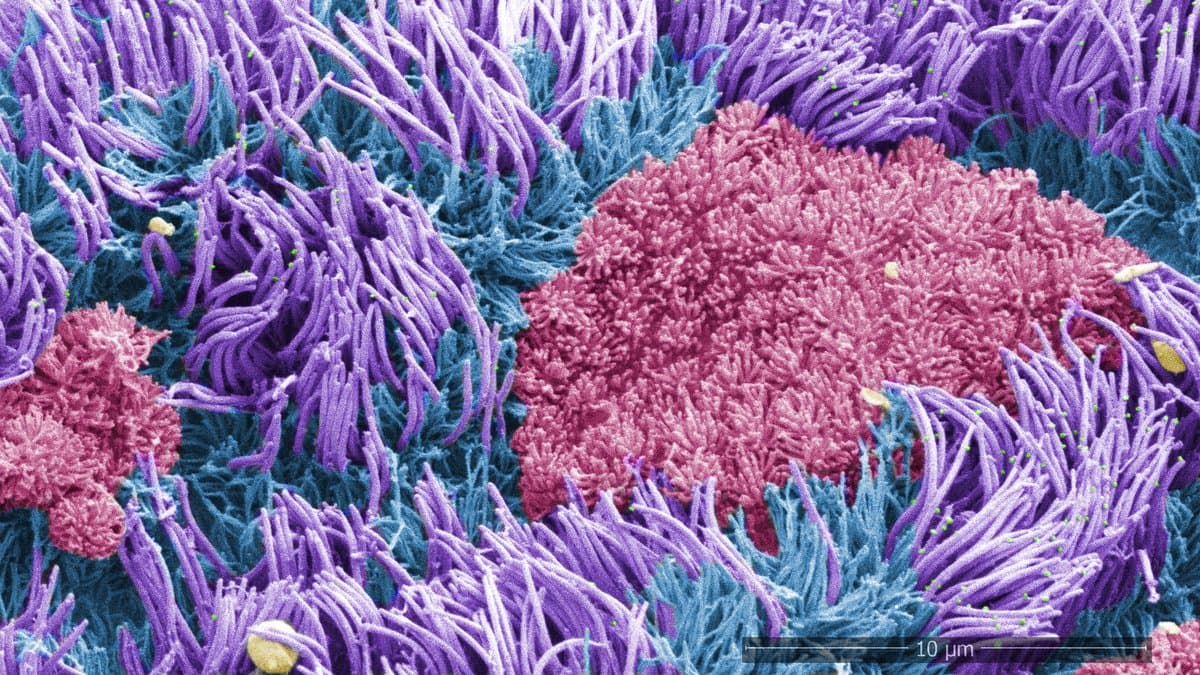
The airways clean themselves. A thin film of mucus catches the dust, smoke and bacteria that arrive with every breath, and millions of microscopic hairs, called cilia, beat in unison to sweep that film up toward the throat, where it is swallowed or coughed away. In one rare inherited disease, the sweeping never works, the film stays where it is, and infection follows.
That disease is primary ciliary dyskinesia. Harvard Medical School, which led the new work, sets out the basics: it is usually diagnosed in childhood, and a child generally has to inherit a broken copy of the same gene from each parent. Changes in any of more than 50 genes can cause it. In about half of cases, one or more organs end up in the wrong place or never form completely. Treatment can slow the disease, but no more. And an estimated 20 to 30 percent of patients reach the end of genetic testing with no answer at all: their cilia plainly do not work, and no known gene says why.
Two of those missing answers surfaced on September 10, 2026, in a paper in Science built on a part of the cilium that nobody had managed to look at properly inside a human cell.
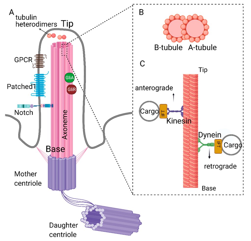
Every cilium is anchored in the cell by a short stretch called the transition zone, the doorway between the body of the cell and the hair standing out of it. Its established job is to decide which proteins get into the cilium and which stay out. Its own structure, and the list of proteins that build it, were another matter: poorly mapped and barely studied at all in the beating cilia of the human airway.
Looking at it meant not taking it apart. By Harvard's account, Haixia Zhou, a research fellow in Alan Brown's laboratory at the medical school, grew cell cultures from the lining of the human respiratory tract and flash-froze them. A beam of ions then shaved each frozen block down until it was thin enough for an electron microscope to see through. Tilted images of what was left were reconstructed into a three-dimensional model of the transition zone sitting where it belongs, inside an intact cell.
That model resolved the paired tubes running the length of a cilium, the doublet microtubules, to better than a nanometer where they pass through the transition zone. With help from AI tools, it also yielded the identities of nine proteins stacked around them. Four of the nine form the complexes that link neighboring pairs of tubes to each other. Two of those four make the major links, and they are the products of two genes: ECT2L and DZANK1.
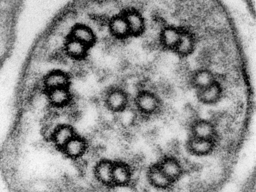
Working with Heymut Omran's group at University Children's Hospital Münster, where Omran also sees children with these diseases as a pediatrician, the team found that people who inherit two broken copies of either gene have primary ciliary dyskinesia. Where either protein was missing, the transition zone lost its shape and the microtubules came out malformed. The cilia grew abnormal bulbous tips, and the mucus stopped moving.
The transition zone was not new to medicine. Faults in it were already known to cause inherited disease, but in the other kind of cilium: the single, nonbeating ones that many cells carry as antennae rather than as oars. The mutations behind primary ciliary dyskinesia had always been found in other parts of the hair. A beating cilium failing because of a fault at its gate is what the paper adds, and the picture it rests on is the first image of the human transition zone taken in place.
Why losing a linker should stop a cilium from beating is not settled. The researchers suspect the failure is one of gatekeeping: with the links between the tubes gone, the transition zone lets the wrong proteins into the cilium and keeps the right ones out. That is their proposed explanation rather than a result. What the paper establishes is the genetic link between a damaged transition zone and a disease of beating cilia.
"This is an example of how combining in situ structural biology with genetics can address fundamental questions and illuminate the causes of human disease," said Alan Brown, co-senior author on the paper and professor of biological chemistry and molecular pharmacology in the Blavatnik Institute at Harvard Medical School.
For families, what this buys today is a name rather than a treatment. What two more genes could do, once laboratories start looking for them, is close part of the gap and turn a blank test result into an explanation. It also hands researchers two more proteins to aim at.
The code the team wrote to build the structure is posted publicly on Zenodo, so another group can rebuild it. And cilia are not only a lung story. The same beating hairs line the inner ear and the reproductive tract, and the transition zone sits at the base of every one of them.
Sources
- science.orgPeer-reviewed
- eurekalert.org