Electrical Activity in the Epithelium: Spikes After Injury and Mechanisms of Cell Orientation

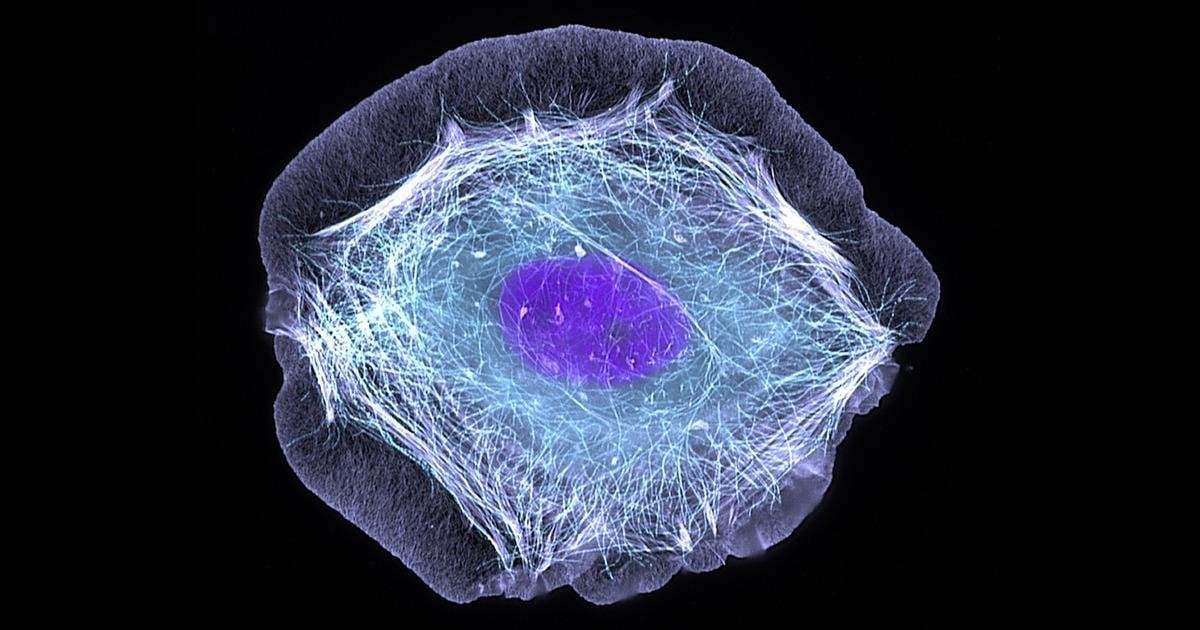
Sun-Min Yu and Steve Granick, researchers at the University of Massachusetts Amherst in the United States, studied how epithelial cells respond to injury. They conducted their experiments on laboratory-grown cell layers, including layers of human keratinocytes, cells of the skin’s outer layer. They grew a continuous layer of cells on a chip containing numerous miniature electrodes and used a laser to injure a small area. The electrodes then recorded repeated changes in electric potential in the fluid surrounding the cells. Some of these spikes were detected several hundred micrometers from the injured area, and electrical activity continued for hours. The researchers published their findings in 2025.
The ability of nerve and muscle cells to generate electrical impulses is well established. The epithelium is more often viewed as a barrier that provides protection and regulates transport, so the appearance of spikes after injury to the cell layer was an unexpected finding in its own right. Yu and Granick suggested that the spikes might help epithelial cells communicate. These spikes were observed in laboratory cultures; their role in living tissue remains to be determined.
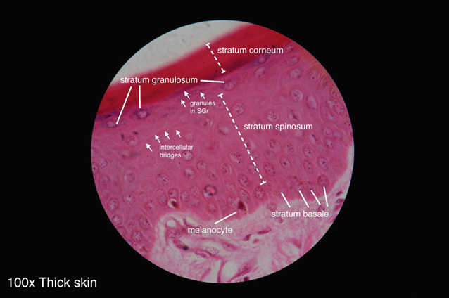
The cells used in Yu and Granick’s experiments included keratinocytes, the main cells of the epidermis, the skin’s outer epithelial layer. In the skin, these cells are tightly connected and continually renewed. Beneath the epidermis lies the dermis, which contains blood vessels, nerves, and connective tissue. Examining keratinocyte membranes and the distribution of ions on either side helps explain these cells’ electrical properties.
Where a Living Cell’s Voltage Comes From
Every cell is surrounded by a membrane made of a double layer of lipid molecules. The fluids inside and outside the cell contain dissolved salts and proteins. Among the dissolved substances are ions, electrically charged atoms and molecules. Their concentrations differ across the membrane. In many cells, the concentration of potassium ions is higher inside than outside, while the concentration of sodium ions is generally lower. Calcium and chloride ions are also distributed unevenly. These differences are maintained by membrane proteins, some of which use chemical energy to transport ions.
One such membrane protein is the sodium-potassium pump. In each cycle, it moves three sodium ions out of the cell and two potassium ions in. This process is powered by the breakdown of one molecule of ATP, a substance that supplies energy for many cellular processes. Other membrane proteins form channels. When a channel opens, specific ions pass through the membrane, driven by electrical forces and differences in concentration. This creates a small separation of charge at the inner and outer surfaces of the membrane. The difference in electric potential across the cell membrane is called the membrane potential. The cell as a whole remains almost electrically neutral: charge separation occurs mainly near the membrane.
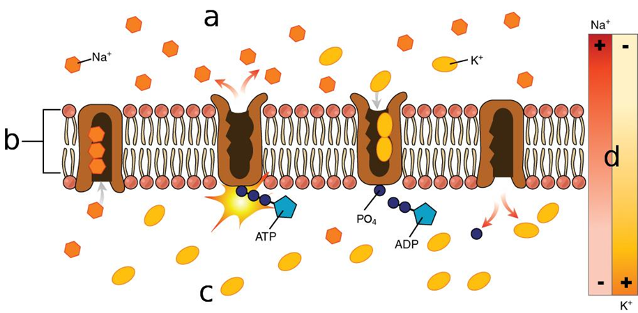
As ion channels open and close, the flow of charged particles across the membrane changes, and the membrane potential may change as well. Redistributing just a small fraction of the ions near the membrane is enough to produce a noticeable change in voltage. The overall composition of the fluids inside and outside the cell remains almost unchanged.
Adjacent epithelial cells form a tightly packed layer that allows ions to pass selectively. Directed ion transport between the two sides of this layer can maintain a voltage known as the transepithelial potential difference. This differs from the membrane potential of an individual cell, which arises between the inner and outer sides of its membrane. In Yu and Granick’s experiments, the electrodes recorded changes in potential in the fluid surrounding the cells.
How a Wound Creates Its Own Electric Field
In the late eighteenth century, Luigi Galvani investigated muscle contractions in dissected frogs subjected to electrical stimulation. In his 1791 publication, he described experiments that later helped lay the foundations of electrophysiology. Alessandro Volta disagreed with him about the origin of the observed effects and the role of metals in contact with one another. In 1800, Volta introduced the first electric battery.
As electrophysiology developed, it became clear that injury to the tissues covering the body changes the distribution of ionic currents. In an intact epithelium, cells maintain a difference in electric potential between its two sides. Injury disrupts this barrier, and ionic currents are redistributed through the surrounding conductive fluid. This creates a directional electric field near the wound. For example, in 2008, Richard Nuccitelli and colleagues, working with skin wounds in mice, measured an electric field of about 177 millivolts per millimeter between the stratum corneum and the epidermis at the wound edge immediately after injury. The field persisted for several days and gradually weakened as healing progressed. Its strength depends on the tissue, the measurement location, and the condition of the wound.
In 2006, Min Zhao and colleagues showed in experiments with cell cultures and laboratory animals that electric fields can guide cell migration during wound healing. They also investigated the intracellular proteins PI3K and PTEN, which are involved in this directional response. Those experiments concerned cell migration in an electric field. In their 2025 study, Yu and Granick examined a different process: the generation of repeated extracellular spikes after injury to a cell layer. They wanted to know how far from the wound the signals could be detected and how they changed over time.
How Electrodes Recorded an Injured Cell Layer’s Electrical Activity
In the 2025 study, Sun-Min Yu grew cultures of human keratinocytes and MDCK cells, an epithelial cell line derived from canine kidney tissue, on a microelectrode array. In each culture, the cells formed a confluent layer. A small area of the layer was injured with a laser, away from the metal electrodes, and changes in potential were then recorded in the fluid surrounding the cells.
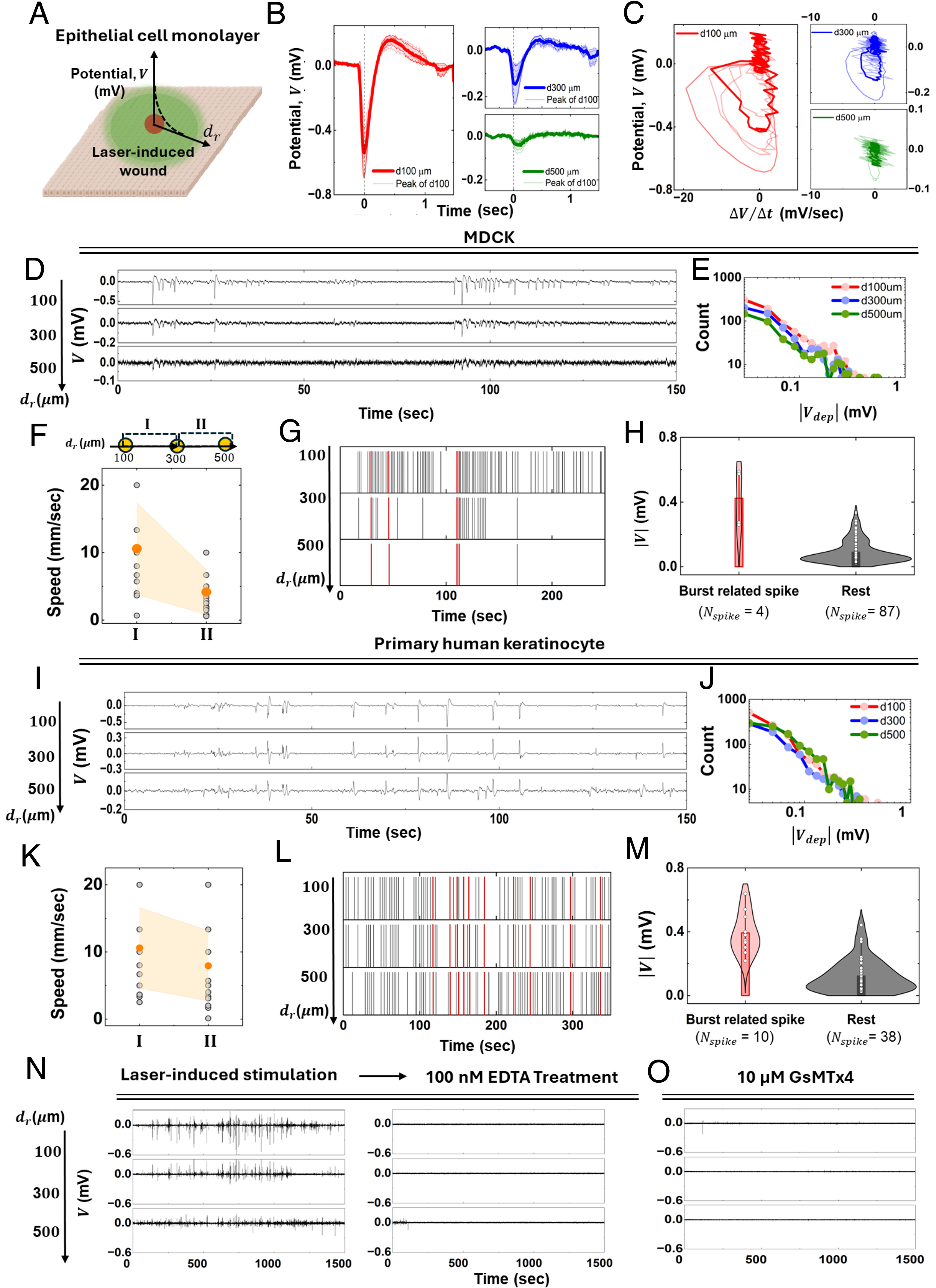
Distinct spikes appeared in the extracellular potential recordings. Many resembled a nerve impulse in shape: the potential first shifted in one direction, returned to baseline, and then briefly shifted in the opposite direction. To describe these phases, the authors used terms from electrophysiology: “depolarization,” “repolarization,” and “hyperpolarization.”
According to Yu and Granick’s findings published in 2025, individual spikes developed over one to two seconds. Some spikes appeared at different electrodes with a time delay. From these delays, the authors estimated an apparent signal propagation speed of about ten millimeters per second on average. Signals were detected hundreds of micrometers from the injury. Recording began approximately ten minutes after laser exposure; in the longest observation, electrical events persisted for five hours. In these experiments, the authors also tested the involvement of mechanosensitive channels: the recorded spikes disappeared after GsMTx4 was applied.
In June 2026, Yu and Granick published a follow-up study of epithelial electrical activity. After laser injury, they recorded four to twelve spikes per minute for more than an hour. According to the authors, the depolarization, repolarization, and hyperpolarization phases developed over one to two seconds. This is about a thousand times as long as typical nerve impulses. Spike frequency and amplitude were comparable at distances of 140 and 740 micrometers from the injury; 740 micrometers was the limit of the range examined. In 2025, the authors had estimated the propagation speed of individual events. The new findings showed that the overall spatial pattern was more complex than a simple wave weakening steadily with distance from the wound. In the same study, Yu and Granick investigated in greater detail the conditions governing the appearance of spikes. They examined the roles of mechanosensitive ion channels, calcium availability, and the cells’ ability to contract.
Calcium, Cell Tension, and Protein Channels
To investigate the factors governing the generation of electrical spikes after injury, Yu and Granick first examined the roles of ions and cellular contraction. Adding EDTA, a substance that binds calcium and other divalent ions, caused the spikes to disappear. However, EDTA can also affect cell-cell contacts, so this result cannot be attributed solely to a change in calcium availability. The researchers then applied blebbistatin, which inhibits myosin II. This protein works with actin to produce cellular contraction. Inhibiting its activity also caused the spikes to disappear.
The next experiments focused on mechanosensitive ion channels, which respond to mechanical changes in the cell. To test their involvement, the researchers used GsMTx4, a substance that affects several such channels. In the 2025 experiments, the recorded spikes disappeared after its application, whereas the 2026 study found partial suppression. Although the extent of the effect differed, both findings pointed to the involvement of mechanosensitive channels in epithelial electrical activity.
The researchers also tested whether these spikes could arise without injury to the cell layer. To do this, they used specific compounds to activate the TRPV4 and Piezo1 channels. Under these conditions, electrical spikes with amplitudes of about 1–10 millivolts appeared even without laser injury.
Yu and Granick’s experiments with ion channels and cellular contraction suggest a possible explanation for why the spikes recur. Injury changes tension in the cell layer, which may alter channel activity and the movement of ions across cell membranes. Cellular contractions, in turn, change the tension again. This feedback could sustain repeated spikes, but further experiments are needed to confirm its role.
How a Cell Chooses a Direction in an Electric Field
Injury to the epithelium can also be accompanied by another electrical phenomenon: the formation of a directional field near the wound. For some motile cells, this field provides a directional cue. Movement in a particular direction in response to such a field is called electrotaxis, or galvanotaxis. In some skin wounds, the center of the injury is electrically negative relative to the surrounding tissue. In the laboratory, electrodes establish the direction of the field. But how does a cell sense this cue and choose which way to move?
In 2026, Nathan Belliveau and his colleagues in the research groups of Julie Theriot, Daniel Cohen, and Michelle Chan sought a protein that helps cells detect the direction of an electric field. They chose the human cell line HL-60 for their experiments. After a special treatment, these cells acquire properties resembling those of neutrophils, motile immune cells.
Belliveau contributed to the major stages of the study, from developing the concept and methods to conducting experiments and preparing the manuscript. Julie Theriot helped develop the concept and provided scientific oversight, while Daniel Cohen and Michelle Chan helped oversee the research and secure funding.
To identify genes that affect cells’ ability to orient themselves in an electric field, the researchers conducted a genetic screen. They reduced the activity of different genes, lowering production of the corresponding proteins, and observed how this affected directed cell migration. Suppressing TMEM154 markedly impaired the cells’ ability to orient themselves in the field. The authors named the protein encoded by this gene Galvanin. Their findings were published in Cell on May 12, 2026.
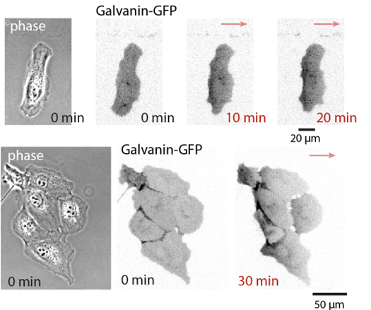
Galvanin is located in the cell membrane, and the portion that extends outside the cell carries a negative electrical charge. In Belliveau and colleagues’ experiments, the electric field caused the protein to move along the membrane toward the positive electrode (anode). The cells under study moved in the opposite direction, toward the negative electrode (cathode). Galvanin therefore accumulated at the rear of the migrating cell. Its redistribution was followed by changes in cellular protrusions and contractions. These observations pointed to a connection between Galvanin’s location and how a migrating cell establishes its front and rear.
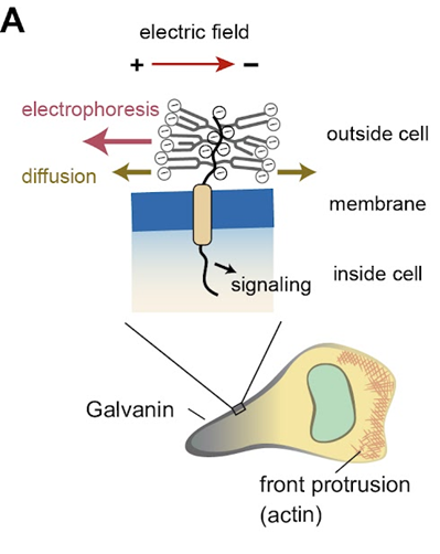
In Belliveau and colleagues’ experiments, cells with impaired Galvanin function continued to move but were less able to orient themselves in the field. Restoring the protein restored directed migration. Experiments with modified versions of the extracellular region demonstrated the importance of its negative charge and confirmed Galvanin’s role in sensing the direction of the field. The researchers have yet to determine the molecular pathway by which the signal from the intracellular portion of the protein reaches the cytoskeleton.
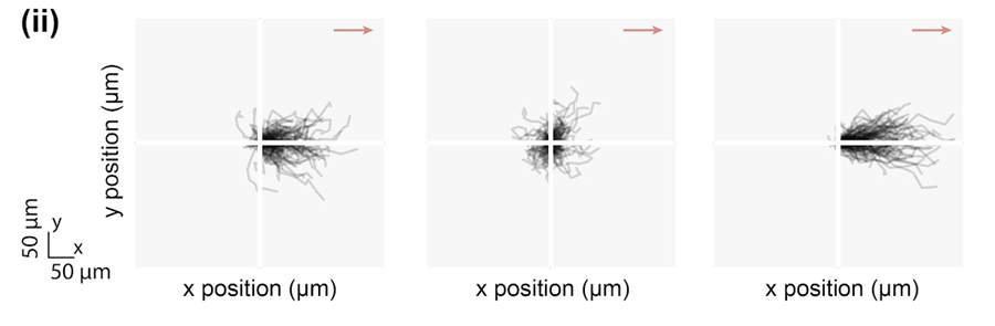
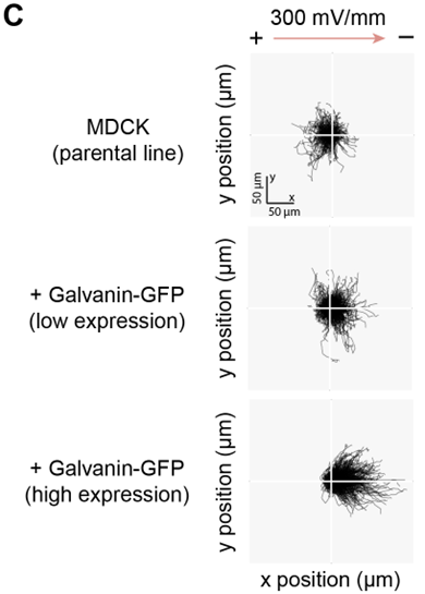
Belliveau and colleagues also studied individual MDCK cells. Without increased Galvanin production, these cells responded weakly to the field and, in these experiments, moved toward the anode. When production of the protein was increased, the cells began moving toward the cathode. However, the behavior of individual cells cannot be directly extrapolated to a confluent layer: MDCK layers can move toward the cathode without this modification.
Two Electrical Phenomena, Different Research Questions
Yu and Granick’s studies and Belliveau’s work explore different manifestations of cells’ electrical properties. Yu and Granick investigated spikes that arise after injury to a confluent epithelial layer and the conditions under which they appear. Belliveau and his colleagues examined how individual motile cells sense the direction of an electric field and the role of Galvanin in this process. Both research teams used MDCK cells, although under different experimental conditions. This raises a new question: could electrical changes after epithelial injury influence directed cell migration through a process involving Galvanin? Such a connection has not yet been established experimentally.
To test this hypothesis, the methods of both studies can be combined. Using the same cell model, researchers would need to simultaneously record electrical spikes, measure the spatial distribution of the electric field, and track both Galvanin redistribution and cell movement. Comparing normal cells with cells in which this protein’s function is impaired would help determine whether Galvanin contributes to their response to electrical changes after injury.
Sources
- Yu S.-M., Granick S. (2025). Electric spiking activity in epithelial cells. PNAS 122(12), e2427123122
- Yu S.-M., Granick S. (2026). Epithelial cells fire voltage spikes. PNAS 123(25), e2601750123
- Belliveau N. M. et al. (2026). Galvanin (TMEM154) is an electric-field sensor for directed cell migration. Cell 189(13), 4107–4121.e22
- Zhao M. et al. (2006). Electrical signals control wound healing through phosphatidylinositol-3-OH kinase-γ and PTEN. Nature 442, 457–460
- Galvani L. (1791). De viribus electricitatis in motu musculari commentarius. Bologna. Digital copy in Smithsonian Libraries. Smithsonian archive
- Bresadola M. (2008). Animal Electricity at the End of the Eighteenth Century: The Many Facets of a Great Scientific Controversy. Journal of the History of the Neurosciences 17, 8–32
- Nuccitelli R. et al. (2008). Imaging the electric field associated with mouse and human skin wounds. Wound Repair and Regeneration 16(3), 432–441
- Belliveau N. M. et al. Galvanin (TMEM154) is an electric-field sensor for directed cell migration. bioRxiv, version 2, August 17, 2025. Source of Figures 4–7 in this article; the original panel numbers refer to the preprint, not the published Cell version. License: CC BY 4.0.