Scientists Map the Human Face as It Forms, Cell by Cell
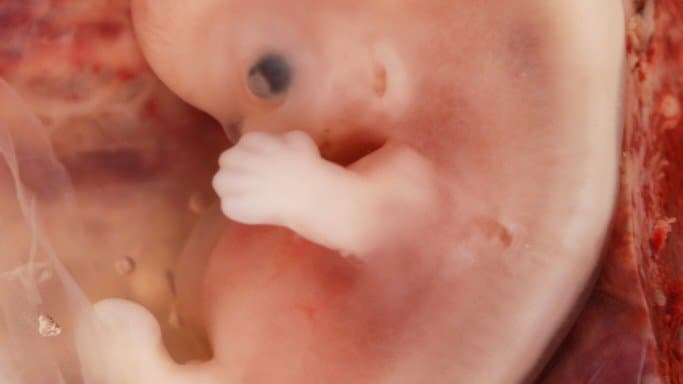
Researchers have mapped the human face as it forms, cell by cell, across embryonic weeks 6 to 11, describing 56 distinct cell states.
The atlas appeared on September 24 in Nature Genetics. The authors offer it as a resource for studying facial development and the birth defects of the face and skull, which they say are among the most common. They also say the cellular steps that link genetic differences to a particular face have remained unclear.
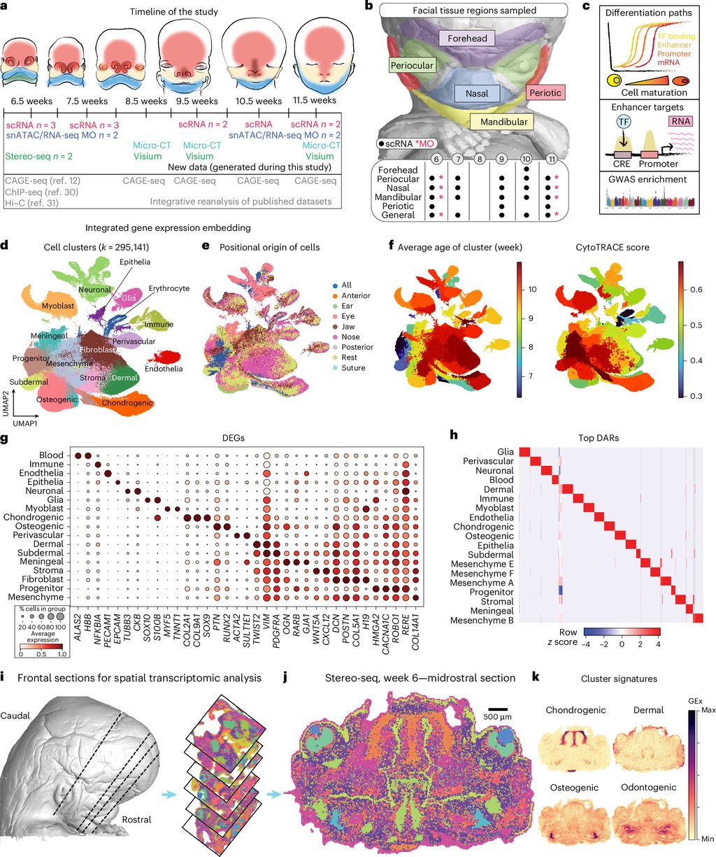
Alek G. Erickson of Stockholm University and Karolinska Institutet, Igor Adameyko of Karolinska Institutet and the Medical University of Vienna, and colleagues combined three measurements on tissue from 20 embryos: which genes each cell was using, which stretches of DNA were open, and where in the tissue each reading came from. The tissue came from elective terminations of pregnancy, donated with consent under approvals from the Swedish Ethical Research Authority.
The team reports a map of 50,000 candidate regulatory regions, the gene switches that set where and when a gene is used, paired with the genes each appears to control. Links between gene activity and facial traits were strongest in the earliest, least specialized progenitor cells and became more region-specific as cells matured.
To test one of those switches, the team used CRISPR to delete a stretch of DNA near the gene PAX1 in mice. The edited mice had smaller bodies, shorter heads and apparent skull defects, which the authors report as evidence that the region matters for normal skeletal development in mice; they add that their analysis cannot show it acts on PAX1 directly.
In a second mouse experiment, embryos bred without cranial sensory nerve clusters had a differently shaped maxilla, the upper jaw, and another bone at the base of the skull, while the cheekbone's length was unchanged. The authors read that as a sign that peripheral nerves fine-tune upper-jaw shape in mammals.
The paper is open access, and the team says the atlas can be browsed online, with the micro-CT scans posted on Zenodo and the raw sequencing files available on request.
Sources
- Nature GeneticsPeer-reviewed