A Transparent Fish Gets a Cell-By-Cell Map of Its Whole Body
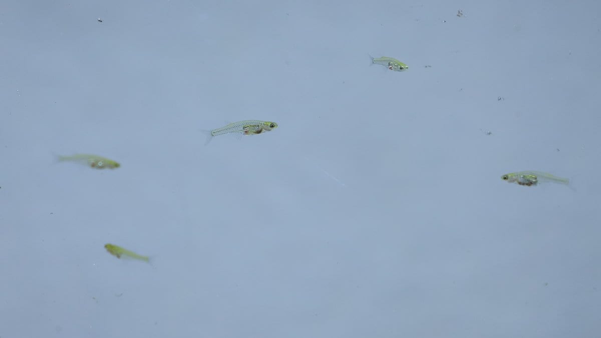
Whitehead Institute researchers have published a map of gene activity in more than 600,000 cells from across the body of an adult fish, the transparent Danionella cerebrum. The institute's release calls it the first such map of a whole adult vertebrate.
The institute describes the atlas as a reference: which cell types the animal has, which genes each uses, and where many of them sit. By its account, researchers studying injury, aging or a drug gain a baseline for the whole animal rather than one organ.
The work was published Oct. 29, 2026, in Science. Kutay Deniz Atabay and colleagues sequenced RNA from cells collected region by region: brain, spinal cord, eyes, organs, fins, head and trunk. They then used spatial methods to put each cell type back where it came from. The fish made that practical: its body is about one centimeter long, and its close relation to the zebrafish means existing genetic tools apply.
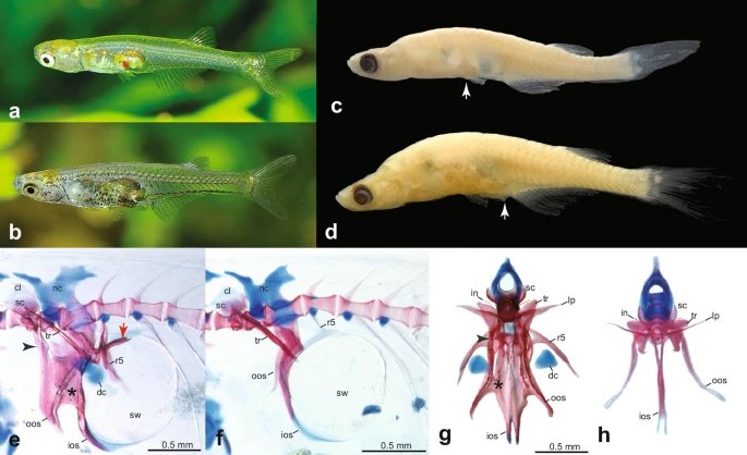
Looking at the whole animal turned up two patterns that are hard to see when tissues are studied separately. Neurons from the same region of the nervous system often had more similar gene activity than neurons elsewhere that used the same signaling chemical. Fibroblasts, the cells of connective tissue, kept region-specific activity of genes that lay out the body during development: cells in the pectoral and pelvic fins held programs tied to forelimb and hindlimb development in other vertebrates. The institute's release says that what those signals do in an adult fish is not known.
The team also injured the telencephalon, part of the forebrain, and tracked gene activity as the missing neurons regrew, from the wound and immune response to new neurons maturing.
"Once you have a spatially resolved atlas, you know what cell types exist, but you also know the neighbors of those cell types in this context," says Atabay, who led the work as a postdoctoral researcher in Peter Reddien's Whitehead Institute lab.
The atlas is public through DigiFish, an interactive resource the team built for browsing cell types and gene activity in the animal.
Sources
- SciencePeer-reviewed
- wi.mit.edu