The Brain Begins Mapping the Visual World Before It Has Seen Anything

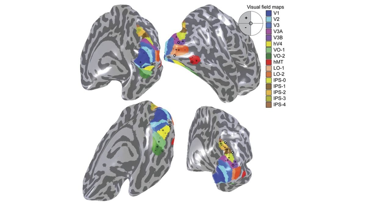
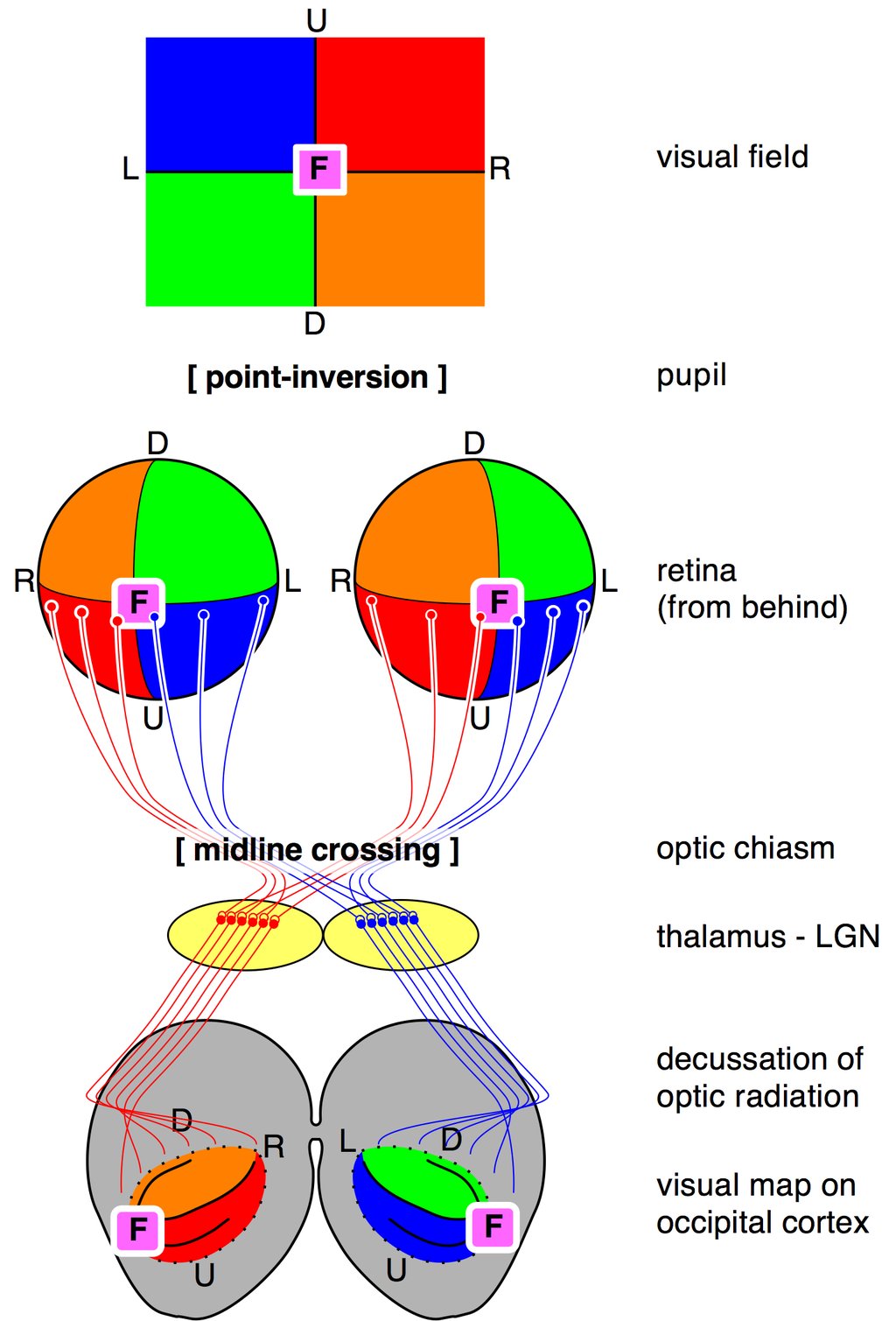
Fix your eyes on a single word on this page. The letters at the center of your gaze are handled by one patch of tissue at the back of your head, the clutter out at the edge of your vision by another, and points that sit side by side on your retina are worked on by cells that sit side by side in your cortex. That orderly layout is called retinotopy, and it is one of the most dependable facts in human neuroscience. The obvious way to explain it is experience: the eyes open, the world pours in, and the map gets drawn by use. A study published on September 30, 2026 in Science Advances reports that a rough version of the map is already in place long before any of that, in fetuses at 21 weeks of gestation.
It comes from So-Hyeon Yoo, Anne-Sophie Kieslinger and Michael A. Skeide at the Max Planck Institute for Human Cognitive and Brain Sciences in Leipzig, with Skeide also at the Christian-Albrechts-Universität zu Kiel. Nobody was scanned for it. The team pooled 871 functional MRI datasets that already existed, counted across every age band from fetal to young adult. All of them were recorded at rest, with nothing shown to anyone. The prenatal and newborn scans came from the Developing Human Connectome Project, the adolescent and adult scans from the Human Connectome Project in Development. Both are open collections, and the group's analysis code and derived data are posted alongside the paper.
Working from resting data means working without a stimulus. In an adult, retinotopy is normally measured by showing someone a moving pattern and recording which parts of the cortex follow it. That is not an option in the womb. Instead, the team used population connective field modeling, which looks for the map in the brain's own slow background activity: for each small patch of one visual area, it estimates which patch of a neighboring area that activity most closely tracks. If those relationships line up in an orderly way, a map is present, whether or not anything has ever been looked at.
Two properties of a visual map survive that treatment. One is eccentricity, how far a point lies from the center of gaze. The other is polar angle, its direction around that center, as on a clock face. Yoo and colleagues report organized maps of both in V2 and V3, the second and third visual areas of the cortex, as early as the 21st week of gestation. This falls in the second trimester of pregnancy, before any visual experience.
They call the maps proto-retinotopic, and the prefix is doing real work. What is there in the second trimester is a scaffold with roughly the right geometry, not the fine-grained adult map. The result is also a statement about order in time: the layout comes first. That is not the same as showing that later experience is unnecessary to finish it.
How much of the visual system comes built in, and how much is assembled by use, is one of the oldest arguments in the field. Evidence about its earliest stages has mostly come from animals and from newborns. An early, rough version of the same organization was described in macaques at birth in eLife in 2017, and retinotopic organization was mapped in human infants in Neuron in 2021. Both are after birth. Carrying the observation back into the second trimester is the new part, and it puts the drawing of the map before the point at which experience could contribute anything.
If the geometry is already laid down in the second trimester, the job of early visual experience looks less like drawing a map than like sharpening one that is handed over at birth. That leaves the harder question standing: what draws it, and what the visual system is using in place of a world it has not yet seen.
Sources
- Science AdvancesPeer-reviewed