Their Memory Scores Looked Alike. Their Brain Scans Did Not

A small number of people in their late seventies and eighties do not lose their memories on schedule. On tests of recall they outscore the average middle-aged adult, decades their junior. The field calls them SuperAgers. In this study the label covers adults aged 75 and over who beat that middle-aged average on memory and hold their own against their age group on a test of planning and attention. That is a looser bar than the age-80 threshold used by the group that coined the term. The usual way to study such people is to sit them beside ordinary older adults and look for the difference.
A team led from Stanford University School of Medicine ran the comparison the other way around. Rather than treat ordinary aging as the reference point and exceptional memory as the thing to be explained, Kanhao Zhao, Yu Zhang and colleagues anchored on both extremes at once: exceptional late-life memory at one end, Alzheimer's disease at the other. Everyone else should fall somewhere on the line between. The authors call the design an "inverted" framework, one that "anchors discovery at the extremes of the cognitive aging spectrum," and it was published Nov. 4, 2026, in Science Advances.
What the scan was trained to see
The scale is built from resting-state functional MRI, a scan of the idle brain. While a person lies still and thinks about nothing in particular, it records how activity in distant regions rises and falls in step, giving a few thousand measures of coordination for each participant. Zhao and colleagues took 180 SuperAgers and 110 age-matched Alzheimer's patients from two long-running studies, OASIS-3 and the Harvard Aging Brain Study, and trained a machine-learning classifier to tell the groups apart from those measures alone. What it settled on, a map of which connections run strong in which group, they named the Alzheimer's Resilience-Vulnerability Connectome, or ARC.
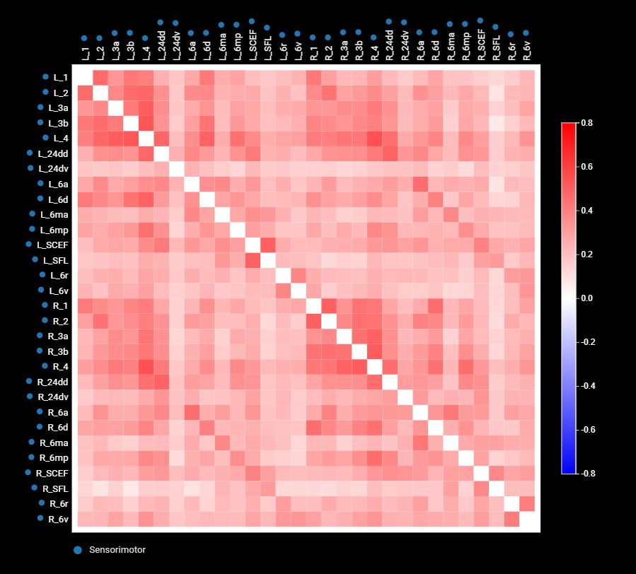
Inside that discovery group the classifier separated the two with an area under the curve of 0.85, on a scale where 0.5 is a coin toss and 1.0 is perfect. That figure comes from holding back a tenth of the data at a time and testing on it, which measures the fit to the material the model was built from. The models were then applied unchanged to 143 people from a third study, the Alzheimer's Disease Neuroimaging Initiative, scanned on different machines and selected with a different memory test. There the score was 0.72, with accuracy, sensitivity and specificity all at 0.71. That second number is the one that says how the signature behaves on data it has never seen.
A split inside a group with no symptoms
The group that mattered was the one in the middle. The team ran the finished classifier over 1,315 older adults who had no cognitive symptoms at all, drawn from the same three studies, and let it push each person toward one pole or the other. About a quarter came out AD-like in each cohort, the rest SA-like. On the standard batteries of memory, attention and daily function, the two subtypes were hard to tell apart at the start.
Hard to tell apart is not the same as identical. The authors report that "age was the only variable that significantly differentiated the two subtypes across both cohorts, with SA-like individuals being younger than their AD-like counterparts," a gap that could on its own produce a difference in later decline. So they pulled it apart: separating each person's age at the first visit from the years that passed afterward left the subtype effect standing in the replication cohort, both on the overall clinical rating and on a timed test of attention and switching. Levels of amyloid and tau, the two proteins that build up in Alzheimer's disease, did not differ between the subtypes at the start.
What separated them was what came later. In both cohorts the clinical ratings of the AD-like group worsened faster and their timed test scores fell faster. More of them went on to a diagnosis of mild cognitive impairment or Alzheimer's, significantly more in the discovery cohort and again, by a narrower margin, in the replication one. The protein scans split the story in two. Amyloid accumulated faster in the AD-like group, most clearly in the frontal and temporal lobes. Tau did not: its accumulation showed no significant difference between the subtypes. That ordering matches the sequence Alzheimer's pathology usually follows, with amyloid spreading early and widely and tau arriving later and in fewer places.
What it would take to call this a test
ARC is a score for a group, derived and checked on research cohorts, and the authors' own summing-up stops where their evidence does. The findings "support the potential utility of ARC for early risk stratification" among older adults without symptoms, they write, "although prospective validation against established and emerging AD biomarkers will be required to establish its clinical utility." The design is observational, which is why they add that it "does not establish specificity to resilience": ARC may be marking a brain that withstands damage, or one that is simply taking on less of it. What they ask for is larger cohorts with biomarker data and "longer longitudinal follow-up."
One further measurement fits the picture. ARC scores tracked a brain's apparent age, estimated from the same scans, more closely than they tracked the person's actual age, and the brains of the AD-like group looked older than their birthdays.
A scan across the whole genome of the ADNI participants turned up three variants near two genes, CLYBL and FRMD6, at a loose threshold the authors set in advance. None cleared the usual bar for a genome-wide finding and none was replicated, and the paper says they "should be considered candidate associations for future validation." That is a long way from the established Alzheimer's risk genes.
All three datasets are open to researchers, and the team has posted its code on Zenodo and GitHub. Another group can rebuild the classifier and run it against a cohort of its own, which is what the authors are asking for.
Sources
- Science AdvancesPeer-reviewed
- europepmc.org