Watch a Flu Virus Make a Cell's Own Droplets Vanish

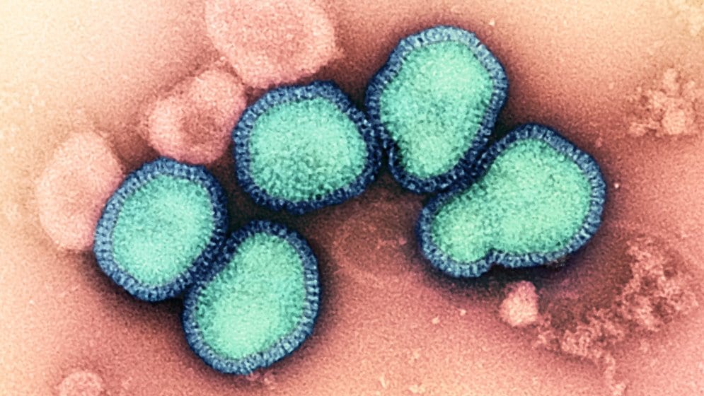
Inside the nucleus of a healthy human cell sit a handful of droplets so small they were only named this century. They have no membrane. They condense out of the surrounding soup the way oil beads in water, holding a cast of RNA-binding proteins in one place, and biologists have come to suspect they help the cell tune which genes it reads and how it braces against stress. They are called paraspeckles. Under a microscope trained on a living cell, they are steady, reliable specks.
Then the flu arrives, and the specks begin to disappear.
That vanishing act is the centerpiece of a study published July 20 in Nature Microbiology by a team at EMBL Hamburg and the Leibniz Research Institute for Molecular Pharmacology in Berlin, with collaborators at Charité–Universitätsmedizin Berlin. The researchers infected human cells with influenza A and then did something that until recently was very hard to do well: they mapped, inside the intact cell, which proteins were physically touching which others, moment by moment, as the infection took hold.
"Watching these tiny organelles in the nucleus dissolve, consistently across every cell line and every flu strain we tested, told us this isn't a side effect of infection," said lead author Iuliia Kotova, in comments reported alongside the paper. "It might be a strategy."
Reading the contacts inside a living cell
The method is worth pausing on, because it is what lets the team make a claim about the real thing rather than a stand-in. Most maps of virus-host interaction come from grinding cells open and fishing out proteins that stick together, which tells you what can bind but strips away the context of the crowded, structured cell. Kotova and her colleagues instead used cross-linking mass spectrometry: a chemical reagent that snaps shut like a molecular staple wherever two proteins are close enough to touch, freezing those contacts in place while the cell is still intact. Feeding the pattern of staples into a modified version of the AlphaFold structure-prediction system, they could then work out not just that two proteins met, but how they fit together.
The payoff was a snapshot of the infected cell's wiring in something close to its native state. Two hijackings stood out. In one, the virus routes its surface protein haemagglutinin through the cell's own internal delivery system, leaning on host proteins to fold and finish it. In the other, the nuclear paraspeckles came apart.
Why a virus would want the droplets gone
Paraspeckles are, in effect, storage. They sequester RNA-binding proteins that the cell uses for its own purposes. Dissolve the droplet, and you release its contents into the nucleus, where an opportunistic virus can put those same proteins to work copying its genome. The team traced the disassembly to specific viral actions: the influenza proteins NP and NS1 interacting with the machinery, the activity of a viral factor called PA-X, and a shutdown of the host enzyme that transcribes genes into RNA. Those are the levers, and pulling them takes the droplets apart.
There may be a second prize in it for the virus. Paraspeckles have been linked to the cell's stress response and to switching on antiviral genes, so scattering them could blunt part of the defense at the same time it hands over raw material. The study stops short of nailing that down, and the authors frame it as a plausible bonus rather than a settled result.
What gives the "strategy" reading its weight is the consistency. The dissolution showed up in every cell line and every influenza strain the team tried. A quirk of one lab dish tends not to survive that kind of repetition; a move the virus reaches for again and again does.
What it changes, and what it doesn't
This is a map, not a medicine. It does not describe a drug, a treatment, or a clinical trial, and nothing here changes how flu is treated this season. Its value is that it names concrete molecular steps the virus depends on. A step the virus cannot skip is, in principle, a step a therapy could block, and the paraspeckle machinery now joins the list of candidates worth probing. Turning any of that into an antiviral is a long road with a high failure rate.
"Our work provides a new way to study flu-host interactions in their native context and with structural insight," said group leader Jan Kosinski. That phrase, native context, is the quiet claim underneath the flashier one. The method is general. The same staple-and-predict approach could be pointed at other viruses in other cells, wherever the question is not what proteins can touch in a test tube, but what they actually do inside a living, infected cell.
The result was peer-reviewed and appears in Nature Microbiology; an earlier version circulated as a preprint on bioRxiv. For now, the most striking thing it leaves you with is an image. Somewhere in a flu patient's airway, in cell after cell, a set of nuclear droplets that were supposed to stay put are quietly coming undone, on the virus's schedule and to the virus's advantage.
Sources
- Nature MicrobiologyPeer-reviewed
- www.embl.org
Medical Disclaimer: This content is provided for general informational and educational purposes only. It is not medical advice and should not be used as a substitute for professional medical advice, diagnosis, or treatment. Always consult a qualified healthcare professional regarding any medical condition, concern, symptom, or treatment decision.