A Standard Way to Isolate the Cells That Train the Immune System
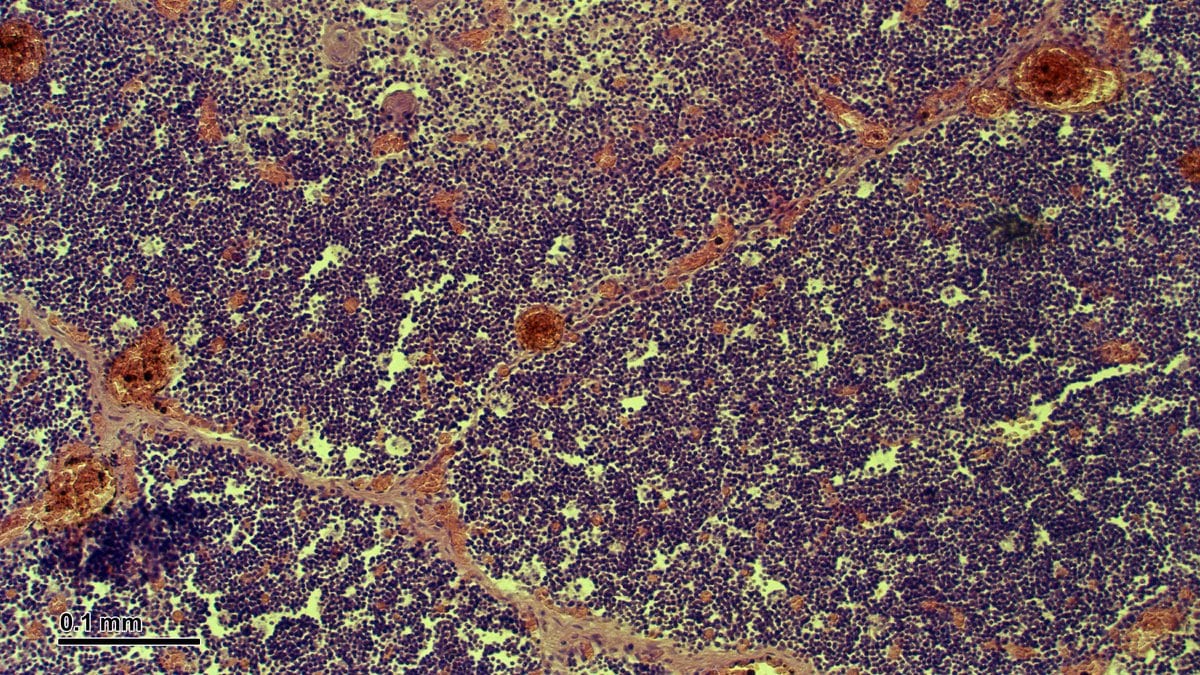
Researchers at Cold Spring Harbor Laboratory have published a set of markers and tissue-handling conditions for separating thymic epithelial cells from the rest of a human thymus. Those are the cells that teach the immune system's T cells what to attack and what to spare.
The authors write that reduced cancer risk, healthy aging and longevity have all been linked to how the thymus works, and that the mechanisms behind those links remain incompletely understood. The paper says consistent tissue processing and analysis are essential before those mechanisms can be studied reproducibly, and it offers "recommendations to improve reproducibility" in work on these cells.
The team found that two signals are enough to tell thymic epithelial cells apart from the other cells in the gland: EpCAM, a protein on the cell surface, plus the cells' own faint glow between 500 and 650 nanometers when a 405-nanometer laser lights them. That combination also separates them from other thymic cells that glow by themselves.
The paper identifies CD205 as the target of CDR2, the antibody classically used to mark thymic epithelial cells in the gland's outer layer. On that basis the authors re-establish CD205 as a reliable marker between those outer cells and the ones in the gland's core, which they confirmed by sequencing gene activity cell by cell.
Alongside the markers, the study compares sample processing and digestion conditions, representative sample size and protein staining patterns. The authors say those comparisons let them reconcile inconsistencies across previously published results. The paper also sets out the smallest set of markers a cell-sorting machine needs to pull out the main thymic epithelial populations and their developmental stages.
The study was carried out by Yong Lin and Hannah V. Meyer of the Simons Center for Quantitative Biology at Cold Spring Harbor Laboratory, together with colleagues. The Journal of Immunology published the work online Nov. 16, 2026.
Sources
- The Journal of ImmunologyPeer-reviewed

Medical Disclaimer: This content is provided for general informational and educational purposes only. It is not medical advice and should not be used as a substitute for professional medical advice, diagnosis, or treatment. Always consult a qualified healthcare professional regarding any medical condition, concern, symptom, or treatment decision.