Self-Destructing Probes Let Biologists See Single RNA Molecules in Living Plant Cells
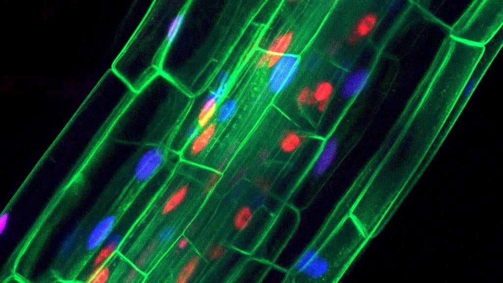
A research group at Sichuan University has built an RNA imaging system for plants in which the fluorescent probe is destroyed unless it is bound to its target, and used it to follow single messenger RNA molecules inside living plant cells. The work was published Oct. 6, 2026, in PLOS Biology, which has posted the article as an uncorrected proof, so figures and wording can still change.
The paper describes the platform as a toolkit for studying plant RNA biology, and the barrier it works around is physical. In a mature plant cell a large central vacuole squeezes the cytoplasm into a thin layer at the edge, which concentrates free probes into a haze bright enough to bury the signal from any one molecule. Animal cells have no such layer, and live single-molecule RNA imaging became routine there first.
Jiayu Zhang, Yiran Tao and colleagues screened plant proteins for a short tail sequence that marks a protein for disposal. They fused the one they found onto a probe built around CRISPR-dCas13X, a gene-editing protein disabled so that it grips RNA without cutting it. Probes stuck to a transcript keep glowing; loose ones are broken down. That raised the contrast between a bright spot and its background by about 6.7-fold, and about 87% of the spots it produced in Arabidopsis thaliana leaves coincided with spots from an established single-molecule staining method.
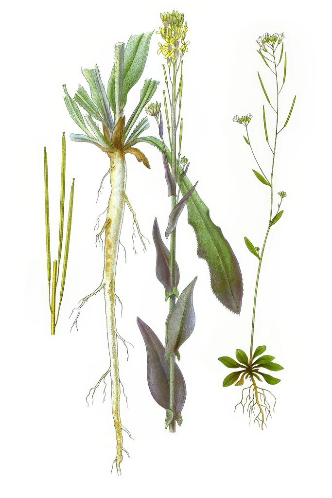
Pointed at FLOWERING LOCUS T, the gene behind a signal that tells a plant to flower, the system caught its messenger RNA waiting roughly 20 seconds at the pores joining one cell to the next, then crossing in about 6 seconds. In plants grafted onto normal roots, the RNA reached a shoot that cannot make it. On roots missing the RNA-binding protein GRP7, far less of it arrived, which the authors report as a contribution of GRP7 to the transport rather than a role specific to this transcript.
Sources
- PLOS BiologyPeer-reviewed