Skip to content
See the World Through ScienceA project of ALLATRA
Source: Peer-reviewedNature Communications1 source

Standing Bacteria Upright on Glass Opened a View of Their Ends
Science

Republish this story

Our work is licensed under Creative Commons BY-NC 4.0. You may republish this piece for free — with credit to ALLATRA Media and a link to the original, unedited beyond length trims, and not for commercial use.

Read the full license

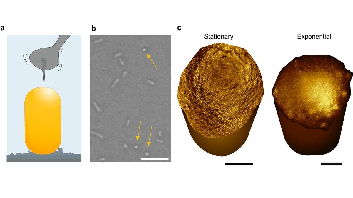
Diagram of an atomic force microscope tip above a rod-shaped bacterium glued to a glass slide, a light-microscope view in which arrows mark round upright cells, and two microscope images of the domed end caps of E. coli.
Panels a to c of the study's Figure 5: the tip tracking a cell held on glass, upright cells seen end-on under a light microscope (yellow arrows), and the exposed poles of a resting and a fast-growing E. coli.Fig. 5 from Anna Scheeder, Joanna Szczepaniak, Yulianna Koziy, Naomi Mara Claro De Oliveira, William Trewby, Renata Kaminska, Jeremy Brown, Colin Kleanthous, Bart W. Hoogenboom (2026), "Nanometer-resolution imaging of living bacteria across species and cellular orientations", Nature Communications — CC BY 4.0, cropped and padded

A team led by University College London has imaged the capped ends of living rod-shaped bacteria at nanometer resolution, after a chemical coating left many of the cells standing on end rather than lying flat. Those ends, called poles, are where a cell has just divided; UCL says they had not been seen at this scale on a live cell before.

The researchers say some bacteria-killing treatments prefer to attack that part of the cell, and that their aim is new leads against drug-resistant strains.
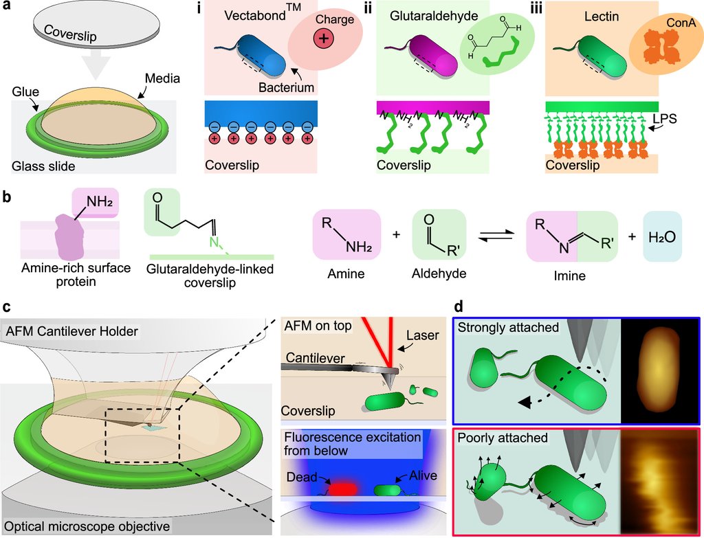
Diagram of three coatings used to stick bacteria to a glass coverslip, the reversible chemical bond formed by glutaraldehyde, and a microscope setup with an atomic force microscope above the slide and an optical microscope below it.
How the cells are held, from the study's Figure 1. Glutaraldehyde forms a bond with proteins on the bacterial surface that water can undo, so a cell stays alive and can still divide while the tip scans it. Fig. 1 from Anna Scheeder, Joanna Szczepaniak, Yulianna Koziy, Naomi Mara Claro De Oliveira, William Trewby, Renata Kaminska, Jeremy Brown, Colin Kleanthous, Bart W. Hoogenboom (2026), "Nanometer-resolution imaging of living bacteria across species and cellular orientations", Nature Communications — CC BY 4.0, resized

The study was published Nov. 9, 2026, in Nature Communications. Lead author Anna Scheeder, of the London Centre for Nanotechnology at UCL, and colleagues coated glass with glutaraldehyde, which bonds to surface proteins and lets go in water. They then ran an atomic force microscope over the stuck cells, its tip a few nanometers wide. Cells settled flat after about 20 minutes; left for about 50 minutes, many stood perpendicular to the glass, a pole facing the probe. Typically at least 90% of the cells kept an intact envelope over two hours, and some grew and divided while being imaged.
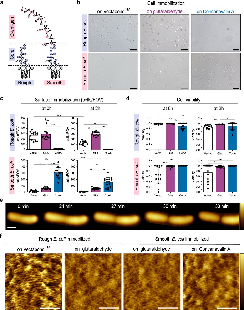
Light-microscope views and charts comparing three glass coatings for holding E. coli, with a row of microscope images following one cell as it grows and splits in two.
Figure 2 of the study compares the three coatings; panel e follows one E. coli over 33 minutes, from a growing rod to two daughter cells, with the tip scanning it throughout. Fig. 2 from Anna Scheeder, Joanna Szczepaniak, Yulianna Koziy, Naomi Mara Claro De Oliveira, William Trewby, Renata Kaminska, Jeremy Brown, Colin Kleanthous, Bart W. Hoogenboom (2026), "Nanometer-resolution imaging of living bacteria across species and cellular orientations", Nature Communications — CC BY 4.0, resized

In E. coli, poles of fast-growing cells carried fold-like irregularities, and the dense network of pore-like features covering the rest of the surface was not resolved. Cells whose growth had stalled had smooth, rounded poles that did carry the network. The authors read the difference as a sign of rebuilding during and just after division.

The coating also held two hard-to-treat species that had resisted this kind of imaging, Pseudomonas aeruginosa and Acinetobacter baumannii. Neither showed E. coli's pore network, which the authors call expected, as both carry far fewer of the proteins behind those features.

Senior author Bart Hoogenboom, at UCL's London Centre for Nanotechnology, said in the university's release: "Imaging the poles of bacteria is like opening a view of the back of the moon. We are looking at something that no one else has seen before."

The work was funded by the Wellcome Trust, the Biotechnology and Biological Sciences Research Council, the Rosetrees Trust and the Engineering and Physical Sciences Research Council.

Sources

By Olga SchmidtEditor-in-Chief, Writer

Spot an error?

Spot an error?

Report an error

Spotted a mistake on this page? Tell us what's wrong and our editors will take a look.

What kind of problem?

Only if you'd like us to be able to follow up. We won't use it for anything else.

We correct mistakes openly. Select any text to flag it. Fixes are logged under our Corrections Policy.

Report an error

Reporting on

Standing Bacteria Upright on Glass Opened a View of Their Ends

What kind of problem?

Only if you'd like us to be able to follow up. We won't use it for anything else.

We read every report. Corrections are logged publicly.