A New AI Tool Lines up Live X-Rays With a Patient's Own Prior Scan
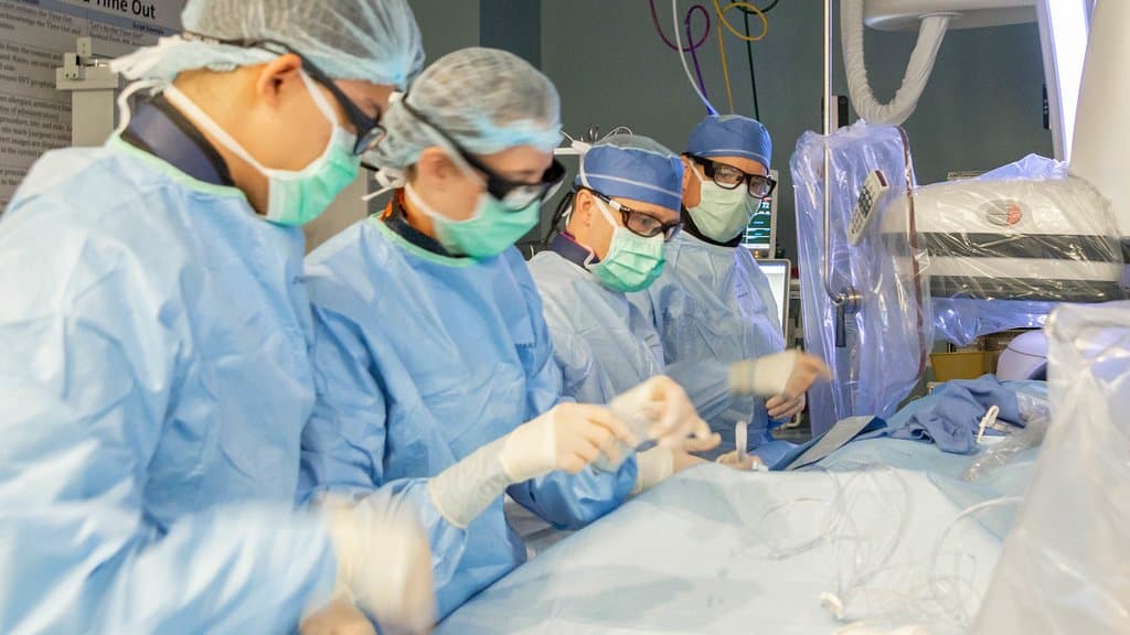
A team at MIT, Harvard Medical School, and several U.S. hospitals has published a method that lines up a live X-ray taken during a procedure with the three-dimensional scan a patient had beforehand, using a small network trained on that patient's own scan instead of on a labeled population.
The method, called xvr, was published in Nature on Sept. 16, 2026. Navigation in image-guided procedures and surgical robotics needs fast and precise alignment between a CT or MRI scan taken before an operation and the flat X-ray images taken during it, the paper says.
Optimizers that work from image brightness have to be tuned by hand for each individual. Deep-learning systems need large sets of hand-labeled data, and they remain limited to the one part of the body they were trained on.
The framework is self-supervised, which means it makes its own training data rather than being given labeled examples. It runs a physics-based simulation of X-ray imaging on the patient's own earlier scan, removing the need for manual annotation. A general model pretrained on thousands of whole-body scans is then adapted to any region of the body with "only 5 min of fine-tuning," and the alignment itself takes seconds.
They describe it as "to our knowledge the largest evaluation of 2D/3D registration on real fluoroscopy to date" (fluoroscopy is live X-ray imaging), and report accuracy across different body structures, scan types, and hospitals, improving on existing methods "by an order of magnitude." What the paper measures is alignment accuracy, not an outcome for patients.
Sources
- nature.comPeer-reviewed
