Skip to content
See the World Through ScienceA project of ALLATRA
Source: Peer-reviewedNature1 source

A Computer Stand-In for Breast Cancer Cells, Built to Predict Drug Responses
AI & Technology

Republish this story

Our work is licensed under Creative Commons BY-NC 4.0. You may republish this piece for free — with credit to ALLATRA Media and a link to the original, unedited beyond length trims, and not for commercial use.

Read the full license

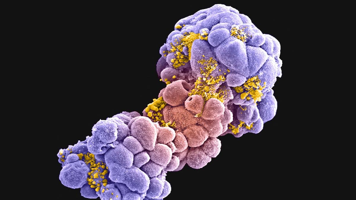
Color-enhanced scanning electron micrograph of a cluster of breast cancer cells treated with the chemotherapy drug doxorubicin, the cells shown in purple and pink with yellow debris on their surfaces.
A cluster of breast cancer cells after treatment with the chemotherapy drug doxorubicin, imaged by scanning electron microscopy and colored afterward. The study measured protein levels in breast cancer cell lines as drugs acted on them. Illustrative image, not from the study."Breast cancer cell spheroid treated with doxorubicin, SEM" (author not named on the source record), via wellcome_collection, CC-BY-4.0 · CC-BY-4.0

Researchers at Westlake University and Peking University have built a computer model of breast cancer cells and trained it on more than 38 million measurements of protein levels recorded as the cells reacted to drugs. Their paper was published in Nature on Sept. 9, 2026.

Rui Sun and Tiannan Guo of Westlake University, Peijie Zhou of Peking University and colleagues call the model ProteinTalks and describe it in the paper as an operational tool for a set of drug-discovery jobs: predicting how well a drug or a drug pair will work, suggesting new combinations, flagging proteins linked to drug resistance, sorting patients by their likely response, and ranking candidate drugs for organoids, which are lab-grown clumps of a patient's own tumor tissue.

To produce that record, the group exposed breast cancer cell lines to drugs and tracked how much of each protein was present at a series of time points. Most existing virtual cell models, the authors write, lack data of this kind.
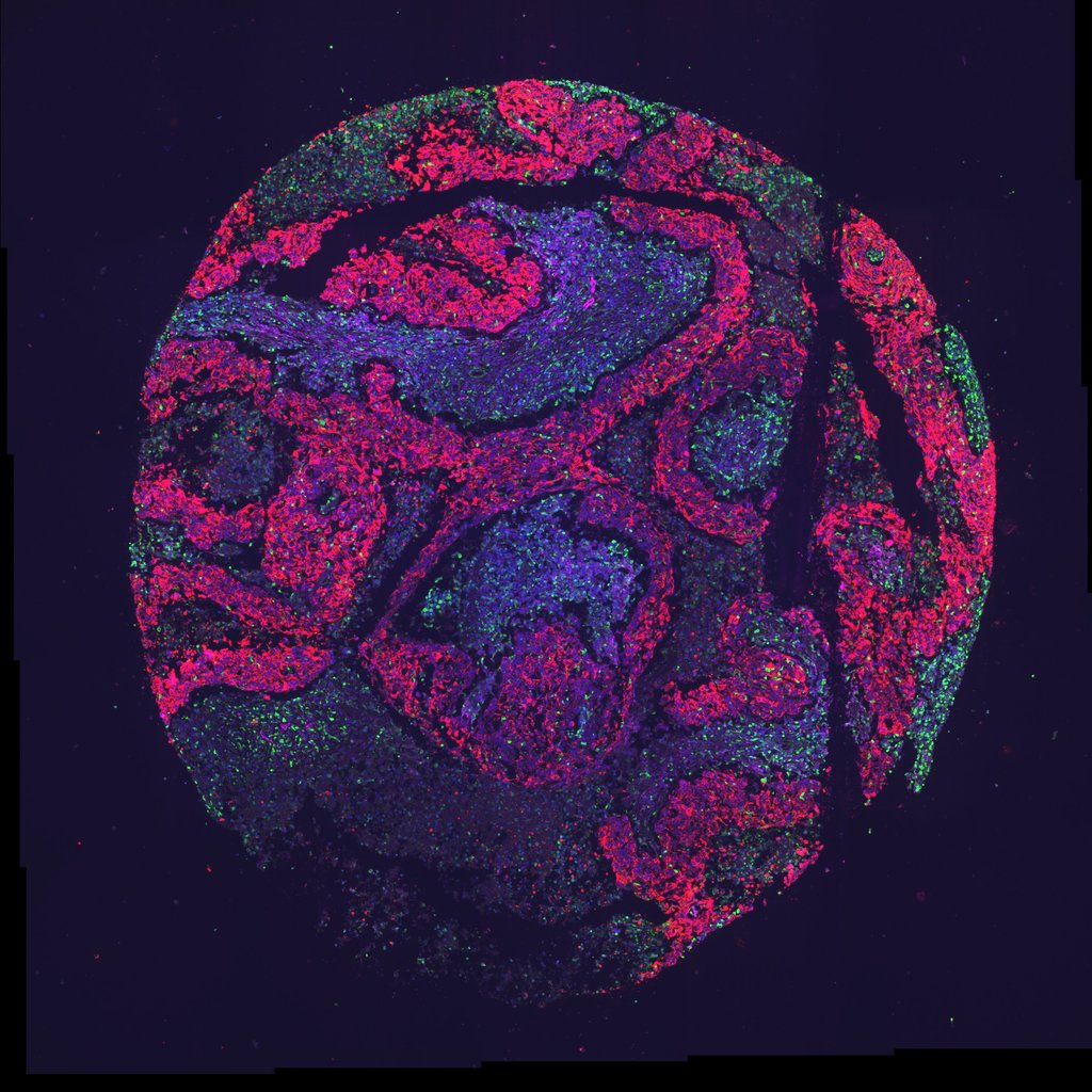
Fluorescence microscopy image of a round core of human breast tumor tissue, with cell populations labeled in pink, purple and green.
Human breast tumor tissue imaged by fluorescence microscopy, with different cell populations labeled by color. The authors report that their model also transfers to biopsy samples taken from patients. Illustrative image, not from the study. — "Image Data Resource - idr0150 - 15148341" by Scherz-Shouval et al, via wikimedia, CC-BY-4.0

The model extends beyond cell lines to organoids and to biopsy samples taken from patients, "generally achieving higher performance than the selected benchmark implementations under the evaluated protocols."

It reports predictions and comparisons rather than patient outcomes, and the study remains at the level of laboratory cell lines, organoid cultures, and biopsy tissue.

The raw measurements have been deposited with the ProteomeXchange Consortium through iProX under accession IPX0007409000, the paper says, and the protein matrix behind the model is available for academic and non-commercial use at db.prottalks.com. The analysis code is on GitHub.

Sources

Spot an error?

Spot an error?

Report an error

Spotted a mistake on this page? Tell us what's wrong and our editors will take a look.

What kind of problem?

Only if you'd like us to be able to follow up. We won't use it for anything else.

We correct mistakes openly. Select any text to flag it. Fixes are logged under our Corrections Policy.

Report an error

Reporting on

A Computer Stand-In for Breast Cancer Cells, Built to Predict Drug Responses

What kind of problem?

Only if you'd like us to be able to follow up. We won't use it for anything else.

We read every report. Corrections are logged publicly.