Cattle Stem Cells Grew Muscle, Nerve and Vessel Cells at Once, With No Assembly Step

Capillaries in living tissue sit a fraction of a millimeter apart, and that spacing is why lab-grown beef has stayed flat. Muscle grown in a dish has no plumbing inside it and starves in the middle, so cultured meat arrives as mince, or as sheets thinner than a millimeter. The standard fix is manufacturing: grow muscle cells in one flask and vessel cells in another, then print, stack or scaffold them into something steak-shaped.
A study published September 2 in Nature Communications goes the other way. Marina Sanaki-Matsumiya, Miki Ebisuya and colleagues at EMBL Barcelona, with collaborators at TU Dresden and UT Southwestern, started from bovine embryonic stem cells and coaxed a single population of them into muscle, nerve and vessel-lining cells at once, then let the cells work out the arrangement themselves.
The route runs through embryology. For two days the stem cells sit in a cocktail that drives them into presomitic mesoderm, the temporary embryonic tissue that skeletal muscle grows out of. That whole population then moves into one muscle-inducing mix of growth factors, and after that nothing is added to make neurons: a fraction of the cells are still poised between muscle and nerve, and the neurons come from them. Adding VEGF, a growth factor that tells cells to build vessels, to the same mix on the same day pulls endothelial cells, the cells that line blood vessels, out of the same source.
Muscle fibers show up in the second week. By the end of the 15-day protocol they carry sarcomeres, the banded contractile units of real muscle, averaging 2.1 micrometers, which is inside the range measured in living cattle. No animal serum is used anywhere in it, and that is not a detail: serum comes from calf blood, and cultured meat that needs it has not left the animal behind.
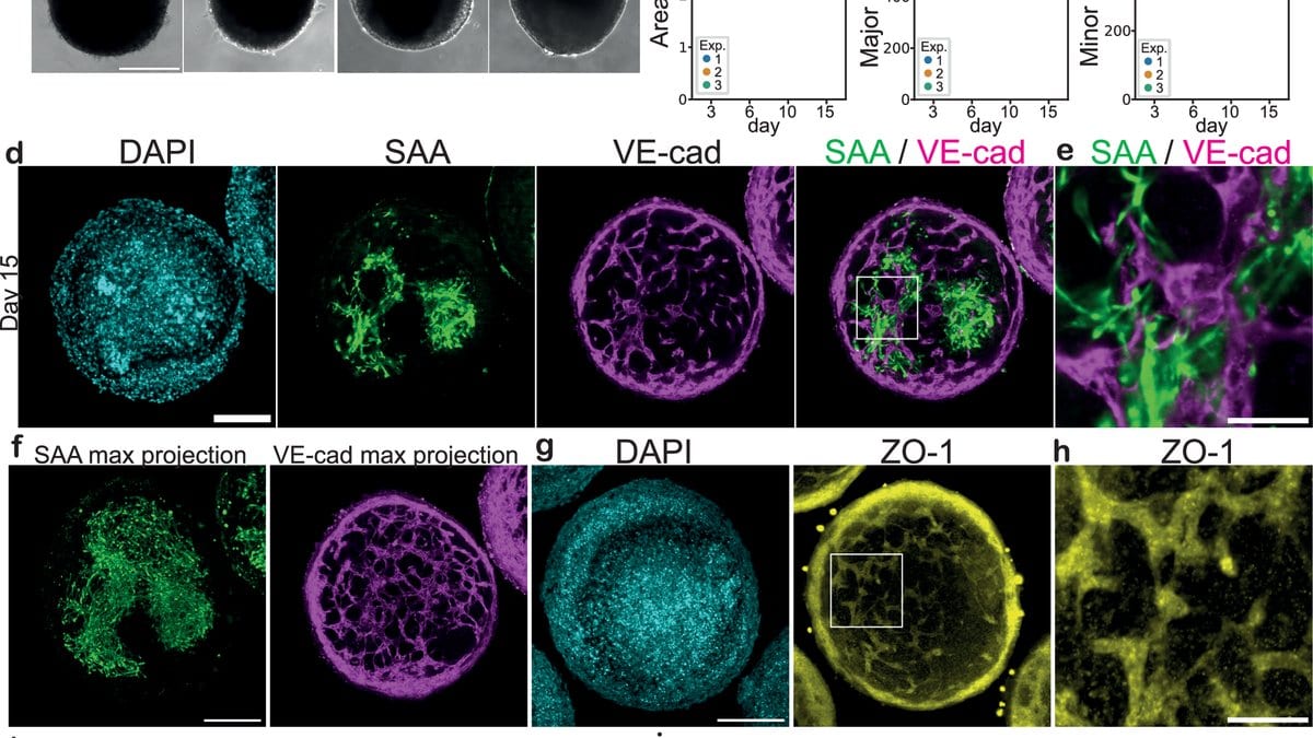
For the three-dimensional version, 20,000 of those cells are packed into a ball and left to turn on a shaker. The aggregates hold their shape at about 600 micrometers across, the size of a coarse grain of sand. That is the object in question, and the authors describe it as a milestone toward self-organizing beef steaks rather than as one.
The muscle in these cultures pulses by itself, calcium rising and falling inside the fibers with nothing prompting it. To find out whether the co-induced neurons were doing the prompting, the team applied curare, the arrow poison that blocks the receptor where a nerve signal lands on a muscle. The pulses were suppressed across 71 matched regions of the same cultures before and after, which is the field's standard demonstration that a nerve is signaling to a muscle.
The evidence runs one way only: the neurons were silenced, never stimulated to drive the muscle, and the contacts between them are rounded and immature, which the authors describe as early junction-like structures rather than finished neuromuscular junctions. Their own verb is that the neurons may transmit signals.
The vessels are the part most likely to be over-read. Endothelial cells emerge on the same timetable and form interconnected, vessel-like networks that run in a regular pattern through the whole aggregate and reach deep inside it, and a stain for a tight-junction protein suggests the cells are sealed to each other the way a vessel wall is. What has not been shown is a lumen, the hollow channel that makes a vessel a pipe, or any flow through one. Vascularized is the authors' own word, and they hedge it themselves: the networks may allow perfusion in the future, and using them for intraluminal perfusion, they write, remains to be seen.
The novelty is real and narrower than it sounds. Muscle and neurons have been grown together from human pluripotent stem cells before, in work this paper cites, and vascularized muscle has been engineered before by preparing the cell types separately and assembling them, or by overriding cell fate with inserted master genes. What is new is the bovine version, and getting the vessel lining out of the same population with no genetic modification and no assembly step. The second half of that is not housekeeping: genetically modified cells are harder to get through food authorization.
On what stands between this and dinner, the authors are unusually frank. One bovine stem-cell line was used, so how the protocol behaves on any other is untested. Matrigel, the gel that keeps the aggregates from falling apart, is derived from mouse tumor cells and would have to be swapped for an animal-free substitute before anyone could eat the result. And they costed it: one aggregate of that size runs 0.39 to 0.99 euros to grow, mostly in growth factors, chemical inhibitors and the Matrigel, and current costs, they write, are commercially unfeasible. Safety and taste the paper does not touch.
The extra cell types have also not paid off yet. Even after 30 days in culture, the team saw no clear positive effect of the endothelial networks or the neurons on the muscle. The whole hope behind co-induction is that nerve signaling will mature the muscle in place of the electrical and mechanical stimulation labs use now, and so far the tissue has the right cells in the right places with no measured improvement to show for them.
The work is peer-reviewed and open access, and it is one laboratory, one cell line and no independent replication so far; a preprint version has been public since 2024. The authors have filed an international patent application on it, PCT/EP2025/054901, and the funding is otherwise public and academic. The sequencing data are deposited at ArrayExpress and the image-analysis code sits in public repositories, so another group can check the analysis rather than take it on trust.
The step the authors call the crucial one is the step they have not taken: connecting the networks inside the aggregate to a vessel system outside it that can be pumped, since it is flow through a vessel that opens its channel in the first place. Until that works, half a millimeter is the ceiling.
Sources
- Nature CommunicationsPeer-reviewed