A Nerve Cell's Branches Turn out to Be Pinched, Not Smooth

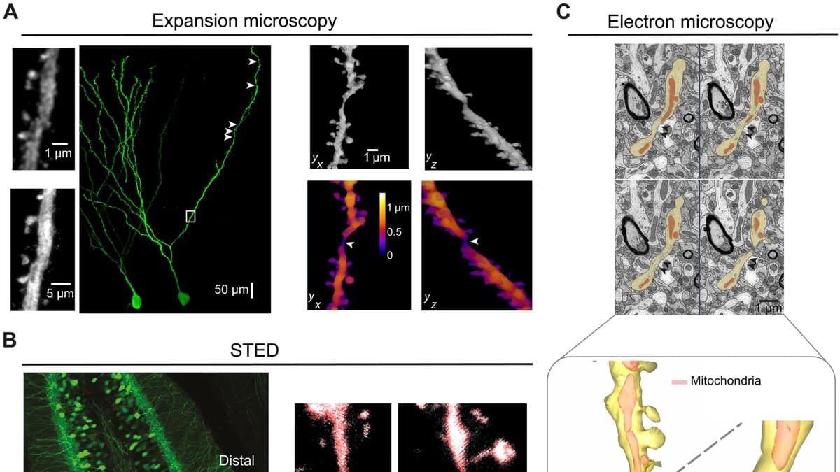
For nearly 150 years the branch of a nerve cell has been drawn as a smooth tube, and there is physics behind the drawing. A dendrite is a neuron's receiving branch, and it tapers as it divides. The taper was read as a design: a cable shaped to carry the small voltages arriving at its far tips down to the cell body with as little loss as possible, splitting at each fork according to a power law published in 1962.
A team led by researchers at the University of Bonn opens its new paper by calling that picture an oversimplification. Run a fine enough microscope along a single branch, and the smooth tube is not smooth. At intervals the shaft narrows by at least half over a stretch a micrometer or two long, then opens out again.
The study, published on September 11, 2026, in Science Advances, calls the narrow points dendritic shaft constrictions. In mice they measure roughly 100 to 500 nanometers across, and they are not spread evenly: they sit on the fine, far-out parts of the tree and are largely absent from the thick branches near the cell body. In the hippocampal cells the team traced end to end, there were about five per branch. The nearest sat some 200 micrometers out from the cell body, which left the far end of the branch divided into a series of walled stretches.
A structure that sat just under the old limit
Why something this common went unrecorded is mostly a question about instruments. That size is at or below what a conventional light microscope can resolve, so a pinch dissolves into a smooth cable. Electron microscopes can see it, but nobody had been looking for it. "Even in high-resolution electron microscope datasets, such local variations in diameter may until now have been regarded as random irregularities rather than biologically meaningful structures," said Tony Kelly, the study's first author, in a statement issued in German by University Hospital Bonn.
Seeing them took four kinds of microscope. Expansion microscopy swells the fixed tissue itself to nearly four times its size, so that structures too close together for ordinary optics move apart. Two super-resolution light microscopes, STED and image scanning microscopy, found the same narrowings in living slices, which answers the obvious worry that the swelling or the chemical fixing had created them. Serial electron microscopy, which photographs tissue slice by slice at far finer detail, found them again.
Then the team went looking in electron-microscope volumes that other groups had already built and put online: a reconstruction of mouse visual cortex from the MICrONS project, and H01, a cubic millimeter of human temporal cortex removed during epilepsy surgery. The pinches are there too, in images the Bonn group did not produce and that anyone can open. The detection code and the data behind the figures are posted publicly as well.
A narrower wire has more resistance
A thinner conductor resists current more, and from the measured shapes alone the team calculates that at a pinch the resistance along one of these dendrites rises more than fivefold. A neuron already uses that trick one scale down. The thin neck of a dendritic spine, the stalk that carries a single synapse, is what keeps each contact its own small electrical room; a constriction would do something similar for a whole length of branch.
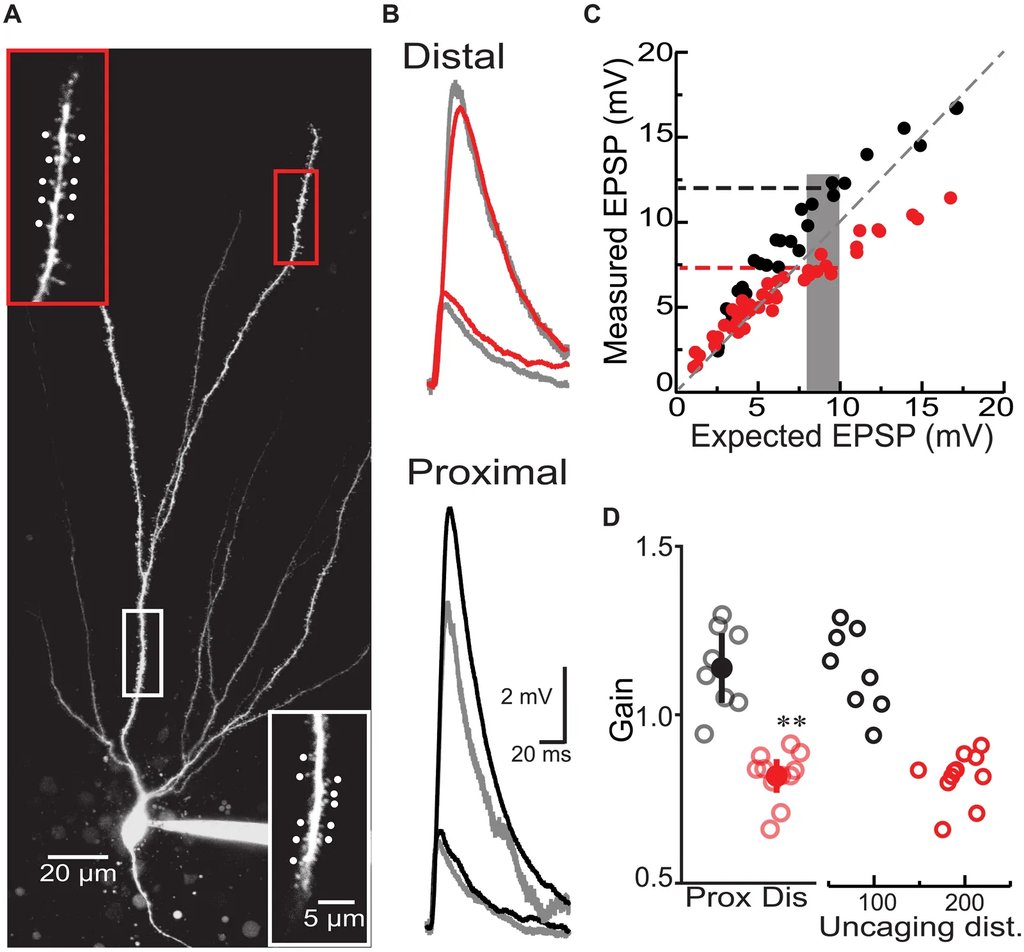
In simulations built on the measured geometry, a signal arriving beyond a pinch grew larger where it landed and reached the cell body at a smaller amplitude. Add more pinches and inputs at the far end stopped adding up straight: several arriving together produced noticeably less than their arithmetic sum.
To test that in tissue, the team used a laser to release glutamate, the signal most synapses use, onto one spine after another along a mouse hippocampal cell. A pipette at the cell body recorded what arrived. Near the base of the branch, the responses added to slightly more than the sum of their parts, a gain of 1.14. Out toward the tip, the same stimulation gave 0.82, which means the far inputs were costing each other. Calcium signals in the outer branches also began at weaker input. Blocking NMDA receptors, the gatekeepers of synaptic strengthening, pushed that threshold back up.
Nobody, though, has recorded at a constriction, and nobody has taken one away. The geometry, the model, and the laser experiments point in the same direction, and the paper says in its own discussion that "a direct causal link between" the constrictions "and these physiological effects remains to be established," for the plain reason that no tool yet exists to alter one on purpose and watch what changes.
The human half is a check, not a second study
The human evidence is thinner than "found in mouse and human neurons" makes it sound. There is no human physiology in the paper at all. The core of it is five neurons re-examined in the H01 volume, where the constrictions were found automatically and, unlike the mouse data, not every one was checked by hand. Two smaller human preparations sit in the supplementary figures: dendrites from epilepsy-surgery slices, and granule cells grown in culture. What that supports is an anatomical claim: that the same structures are there in human tissue. The size range above belongs to mice.
"Our results point to a previously unknown structural feature that could allow individual dendrites to process information locally, rather than merely passing it on," said Heinz Beck, the paper's senior author, whose laboratory at the Institute for Experimental Epileptology and Cognition Research in Bonn led the work. The constrictions could be a new basic element in how neurons carry out complex computations, he added, "although their precise biological role and the way they arise still have to be clarified."
What the constrictions are made of is unknown. Whether they are laid down during development, whether activity changes them, whether they look different in a diseased brain: the paper lists all three among the questions it could not address. It has established a shape, and made a careful case for what the shape should do. The rest waits on a way to take hold of one.
Sources
- Peer-reviewedScience Advances
- doi.org