Skip to content
See the World Through ScienceA project of ALLATRA
Source: Peer-reviewed1 source

Silencing a Rarely Studied Brain Layer Slowed Mouse Brain Waves
Science

Republish this story

Our work is licensed under Creative Commons BY-NC 4.0. You may republish this piece for free — with credit to ALLATRA Media and a link to the original, unedited beyond length trims, and not for commercial use.

Read the full license

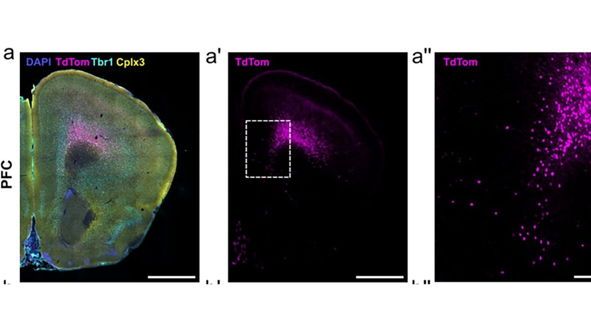
Three-panel fluorescence micrograph of a mouse prefrontal cortex hemisection: magenta-labelled cells form a dense band deep in the cortex at layer 6b, with a magnified close-up at right.
Layer 6b cells (magenta) in a mouse prefrontal cortex section: a stained hemisection, the same section showing only the layer 6b label, and a close-up of the boxed region. Panels a, a′ and a″ of the study's Figure 1.Figure 1 from Elise J Meijer, Marissa Mueller, Lukas B Krone, Tomoko Yamagata, Anna Hoerder-Suabedissen, Sian Wilcox, Hannah Alfonsa, Atreyi Chakrabarty, Luiz Guidi, Peter L Oliver, Vladyslav V Vyazovskiy, Zoltan Molnar (2026), "Cortical layer 6b mediates state-dependent changes in brain activity and effects of orexin on waking and sleep", eLife — CC BY 4.0, cropped · CC-BY-4.0

Mice whose layer 6b neurons had been switched off slept and woke for the same total number of hours as normal mice, but the electrical rhythms running through their brains were slower and weaker, researchers at the University of Oxford report in eLife.

The authors call layer 6b by far the least studied of the cortex's layers. They read the recordings as evidence that it helps set brain state (the difference between waking, deep sleep and dreaming sleep) and that it shapes how the brain responds to orexin, a chemical that promotes wakefulness.

The team made long EEG recordings, which track brain-wave activity through electrodes on the skull, in mice in which a subset of these neurons had been silenced from birth. In those animals, theta waves were slower while the mice were awake and during REM sleep, the dreaming stage. The paper puts that rhythm at 6 to 9 Hz, or cycles per second. Total EEG power fell sharply, most of all during non-REM sleep. The total amount of waking and sleep did not change, and neither did the animals' response to being kept awake.

The group also infused orexin A straight into the brain's fluid-filled ventricles. Both the silenced mice and normal mice spent more time awake afterward, but in recordings taken from the back of the head, the slow waves that mark deep non-REM sleep were then weaker in the silenced animals.

Layer 6b neurons respond directly to orexin and connect widely across the brain, which the authors cite as their reason for studying the layer. The paper, by Elise J. Meijer, Zoltan Molnar and colleagues at Oxford, is open access.

Sources

By Olga SchmidtChief Editor, Writer

Spot an error?

Spot an error?

Report an error

Spotted a mistake on this page? Tell us what's wrong and our editors will take a look.

What kind of problem?

Only if you'd like us to be able to follow up. We won't use it for anything else.

We correct mistakes openly. Select any text to flag it. Fixes are logged under our Corrections Policy.

Report an error

Reporting on

Silencing a Rarely Studied Brain Layer Slowed Mouse Brain Waves

What kind of problem?

Only if you'd like us to be able to follow up. We won't use it for anything else.

We read every report. Corrections are logged publicly.