Anesthesia Leaves the Same Mark on a Worm's Brain and a Human's
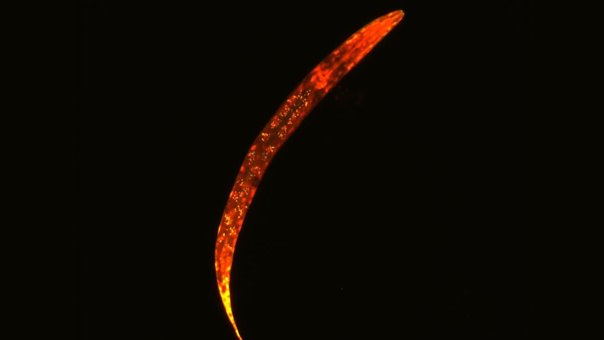
A research team reports that brains as different as a nematode worm's and a person's change in the same measurable way under anesthesia: activity in any one part of the brain keeps less of a trace of what it was just doing, and separate parts fall out of step with one another.
The work, led by Andrea I. Luppi of the University of Oxford and Bratislav Misic of McGill University, was published on September 29, 2026, in Nature Neuroscience, open access. The authors say most anesthesia research looks at one species, one drug and a handful of hand-picked measures at a time, which leaves open the question of what drugs with different molecular targets have in common.
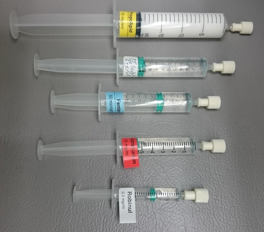
Their compiled dataset covers human, macaque, marmoset, mouse, larval zebrafish and nematode under seven anesthetic regimes, spanning 700 million years of evolution. Rather than pick measures in advance, the team ran every recording through more than 6,000 statistical descriptions of how a signal behaves over time, drawn from the published literature. The shared profile that came out, shorter internal timescales and weaker coupling between regions, held in all six species.
The authors also report running the effect backward. In macaques kept under the anesthetic propofol, electrodes stimulating the centromedian thalamus, a hub deep in the brain, reversed the profile, and the animals responded to their surroundings again. Stimulating a different thalamic target in the same animals did not wake them.
Most of the recordings come from experiments run for earlier studies; the zebrafish data are new to this paper. The profile also lines up with where genes for excitatory and inhibitory signaling, the brain's accelerator and brake, are most active, and a model of how fast signals pass between neurons reproduces it. The paper describes the result as a shared neural endpoint of anesthesia: whatever the drug, local activity is cut off from its own past and from the rest of the brain.
Sources
- Nature NeurosciencePeer-reviewed