Skip to content
See the World Through ScienceA project of ALLATRA
Source: Peer-reviewedNature1 source

Human Brain Proteins Made Mouse Proteins Fold the Same Way
Science

Republish this story

Our work is licensed under Creative Commons BY-NC 4.0. You may republish this piece for free — with credit to ALLATRA Media and a link to the original, unedited beyond length trims, and not for commercial use.

Read the full license

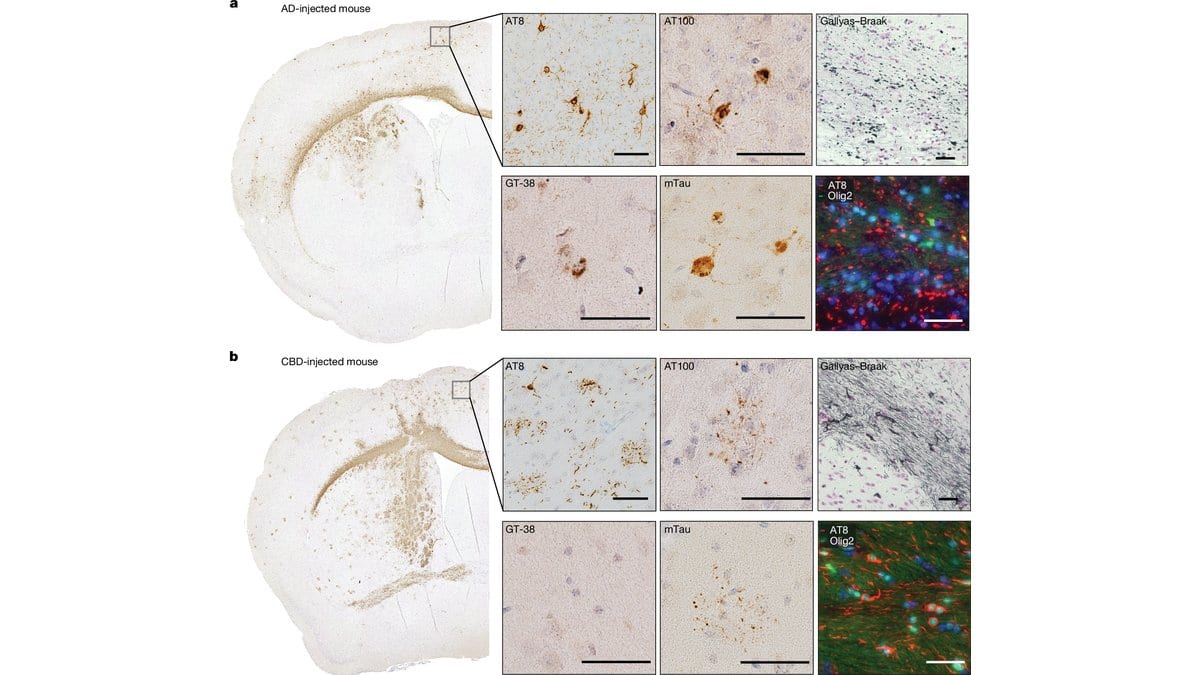
Plate of microscope images: two whole mouse brain sections stained brown, with magnified panels showing dark tau deposits in nerve cells and green and red fluorescence panels.
Mouse brain sections 9 months after injection, stained to show where tau gathered. The upper set, a, comes from a mouse given Alzheimer disease filaments and the lower set, b, from one given corticobasal degeneration filaments.Fig. 1 from Sofia Lövestam, Aki Shimozawa, Airi Tarutani, Reiko Ohtani, Masami Masuda-Suzukake, Kazuko Hasegawa, Andrew C. Robinson, Yuko Saito, Shigeo Murayama, Mari Yoshida, Hisaomi Suzuki, Mitsumoto Onaya, Masato Hasegawa, Michel Goedert, Sjors H. W. Scheres (2026), "Prion-like transmission of human tau strains in the mouse brain", Nature — CC BY 4.0

A British and Japanese team reports that tau filaments taken from the brains of people who had Alzheimer's disease or corticobasal degeneration, injected into the brains of ordinary mice, made the mice's own tau assemble into filaments with the same structures as the injected material. Tau is a protein that clumps inside nerve cells in Alzheimer's disease, the most common of more than 20 brain diseases defined by tau filaments; corticobasal degeneration is a far less common one.

Most neurodegenerative diseases are thought to spread the way prion diseases do, with a misfolded protein forcing its shape onto normal copies of itself. The authors describe one assumption behind that account as a pillar of the prion hypothesis that had not been demonstrated: that a strain keeps its structural identity when it is passed on. They also say the mouse is now a suitable model for studying how distinct tau shapes drive each disease's damage. The paper was published online in Nature on Sept. 30, 2026.

Sofia Lövestam, Michel Goedert and Sjors H. W. Scheres of the MRC Laboratory of Molecular Biology in Cambridge, UK, did the work with colleagues at the Tokyo Metropolitan Institute of Medical Science. The mice were wild type, carrying no added human tau gene, and their brains were examined 9 to 12 months after injection. In mice given Alzheimer's filaments, 83% of those recovered were paired helical filaments, the form found in Alzheimer's brains. In mice given corticobasal degeneration filaments, the structures matched the type I filaments found in that disease. Mouse and human tau sequences are identical in the core of these filaments, which, the authors say, suggests there may be no species barrier.
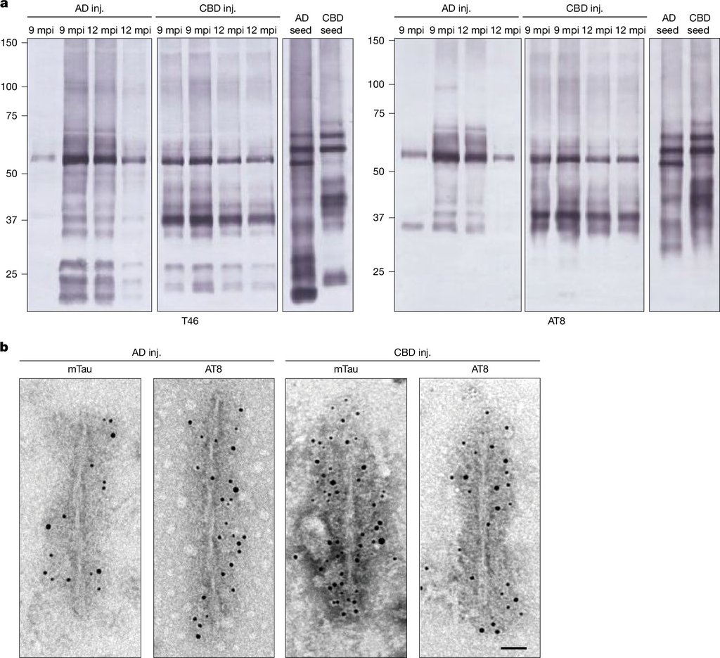
Western blot lanes of insoluble mouse tau beside electron micrographs of filaments studded with dark gold particles.
Insoluble mouse tau builds up 9 and 12 months after injection, and electron microscopy with gold labeled antibodies picks out the filaments it forms. Fig. 2 from Sofia Lövestam, Aki Shimozawa, Airi Tarutani, Reiko Ohtani, Masami Masuda-Suzukake, Kazuko Hasegawa, Andrew C. Robinson, Yuko Saito, Shigeo Murayama, Mari Yoshida, Hisaomi Suzuki, Mitsumoto Onaya, Masato Hasegawa, Michel Goedert, Sjors H. W. Scheres (2026), "Prion-like transmission of human tau strains in the mouse brain", Nature — CC BY 4.0

The pattern of damage tracked the disease the filaments came from. Alzheimer's filaments, the authors report, produced tau deposits confined to nerve cells, while corticobasal degeneration filaments also produced deposits in glial cells, the brain's supporting cells, including the plaque-like inclusions around astrocytes seen in people with that disease. Tau filaments in earlier genetically engineered mouse models had structures unlike those in human brains.

Sources

By Olga SchmidtEditor-in-Chief, Writer

Spot an error?

Spot an error?

Report an error

Spotted a mistake on this page? Tell us what's wrong and our editors will take a look.

What kind of problem?

Only if you'd like us to be able to follow up. We won't use it for anything else.

We correct mistakes openly. Select any text to flag it. Fixes are logged under our Corrections Policy.

Report an error

Reporting on

Human Brain Proteins Made Mouse Proteins Fold the Same Way

What kind of problem?

Only if you'd like us to be able to follow up. We won't use it for anything else.

We read every report. Corrections are logged publicly.