Deep Sleep Sets Brain Fluid Moving. In Older Adults, That Surge Is Mostly Missing

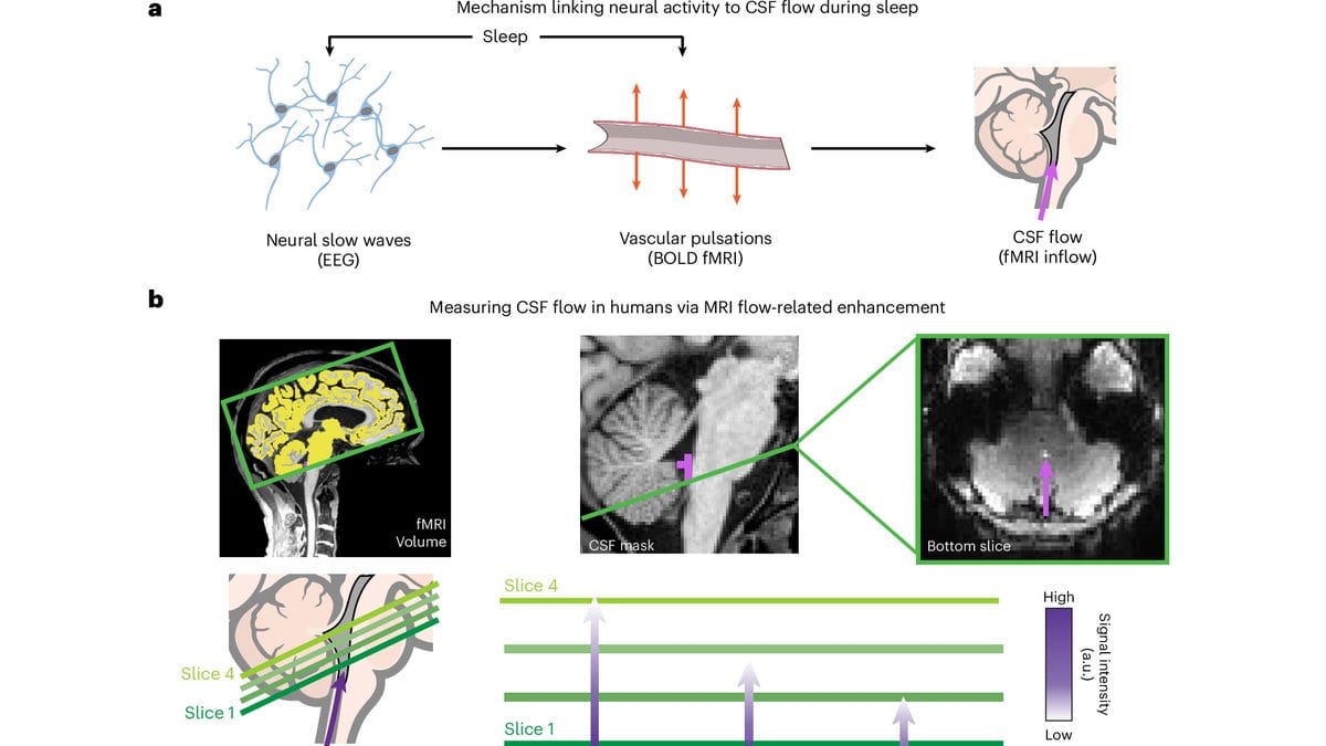
The scans began around midnight. A volunteer in a cap of electrodes lay down in an MRI machine at Boston University or at MIT, and was asked to rest with their eyes closed until sleep came. The machine was positioned so that the bottom edge of the imaged volume cut through a small fluid-filled space at the base of the brain. In a young sleeper, the deep stages of sleep known as non-REM change what the fluid there does: it begins to surge in and out in slow, heavy pulses, roughly one every 20 seconds. Nobody had watched that rhythm during sleep in an older brain.
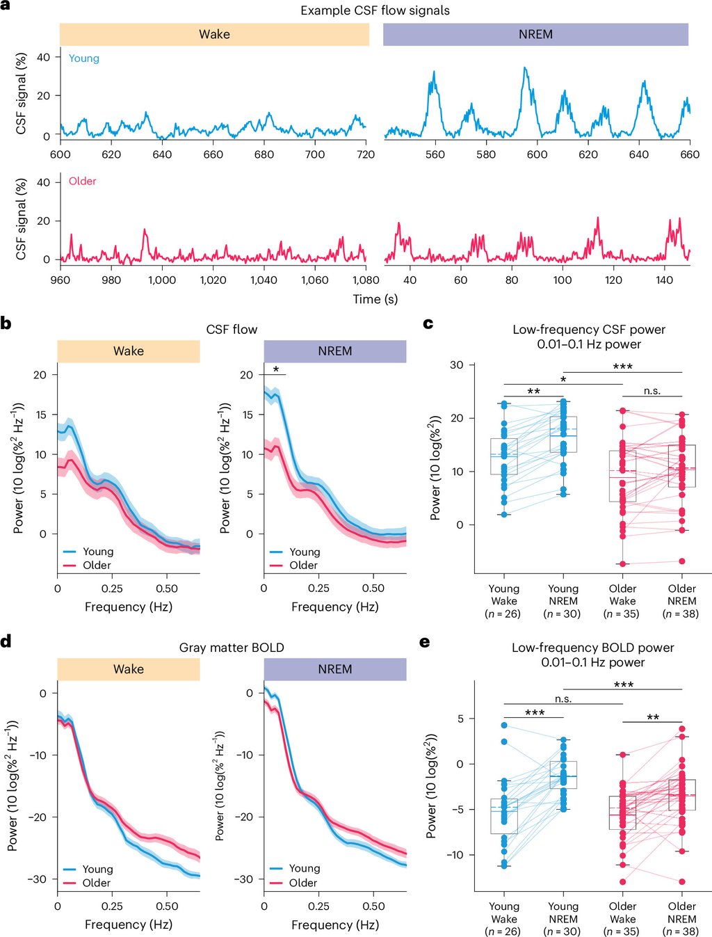
In older adults, it barely happens. Sydney Bailes, Laura Lewis and their colleagues report that the jump in fluid flow from waking into sleep was close to flat in the older group. The study, published Nov. 13, 2026, in Nature Aging. was conducted by researchers at MIT, Boston University, Massachusetts General Hospital’s Martinos Center and Brown University, with Bailes and Lewis among the MIT-affiliated authors. Thirty-one young adults and 41 aged 60 and over remained in the overnight analysis, after exclusions for staying awake throughout or for moving too much. These were not full nights in a sleep laboratory: each person got one to three runs of sleep opportunity, 25 minutes each.
The measure is power in decibels: how much of the fluid signal sits in the slow band the sleep waves occupy. From waking into sleep, the young group gained 3.82 decibels there on average. The older group gained 1.15, a rise that did not reach statistical significance. Flow was already lower in the older group while they were awake, but the gap widened significantly once they slept, which is the finding. The design compares age groups at a single sitting and follows nobody over time, so it says that older adults had much weaker waves, not that any one person's waves weakened with age.
Two ways to lose the same wave
In deep sleep, large populations of neurons fire together in slow electrical waves. Those waves make blood vessels widen and narrow in step, so the volume of blood inside the skull rises and falls. Because the skull cannot expand, fluid moves in as blood leaves and back out as it returns. Both of the first two steps were weaker in the older group. Their sleep slow waves were smaller, which earlier sleep research had already established, and their slow swings in blood volume were smaller too. The signals were also less tightly locked to each other: the correlation between blood volume change and fluid flow was significantly lower in the older adults.
A statistical mediation model put the neural step first: the age-related loss of fluid flow ran through the loss of slow waves, with the weaker blood swings following from that rather than the other way round. Even with both accounted for, age still predicted some of the loss, leaving room for a mechanism the model does not contain.
All of that rests on correlations between signals recorded during sleep. So the team ran a second set of experiments in daylight, with everyone awake and the system driven on purpose. A checkerboard flickering 12 times a second was used to push neural activity. The electrical response it produced was the same in both age groups. But the blood response in the visual cortex was smaller in the older participants, and so was the fluid flow it evoked. Repeated 15-second breath-holds pushed the blood vessels directly, with exhaled carbon dioxide measured through a nasal tube, and the older group's vessels widened less. Those arms used different and partly overlapping samples, and what ties them to the night is a correlation: people whose daytime flow responded weakly also had weaker flow asleep.
One structural measure lined up too. Among the older adults only, those with more gray matter in the frontal lobes had stronger fluid waves during sleep; gray matter elsewhere showed no such relationship. Frontal regions are where sleep slow waves are largest.
What the scanner did not watch
The reason this line of work draws attention is the idea that sleep helps wash waste out of the brain. That is not what was measured here. The scanner recorded how fast fluid moved through one small space at the base of the brain. "The functional consequences of reduced large-scale ventricular CSF flow for waste clearance are still not established, and waste clearance was not measured in this study," the authors write. Cerebrospinal fluid does carry dissolved material out of brain tissue. Whether slower flow means that less of it leaves is untested, and a direct test, with a tracer injected into patients' spinal fluid, is named as future work.
Dementia sits further out still. In a 2020 review in Science, Maiken Nedergaard and Steven Goldman argued that failing fluid clearance could be a final common pathway to dementia. That hypothesis is the backdrop here, and it is not what this study tested. Nobody's memory or thinking was assessed. Cognitive impairment was in fact one of the exclusion criteria, which is also what gives the finding its value: the change shows up in people with no diagnosis. The paper's own claim stops at a hedge: that reduced flow during sleep "could be an early process linked to age-related decline in cognition and brain health."
The waves themselves were not new. Nina Fultz, Lewis and colleagues first recorded them in young sleepers in Science in 2019. An independent team in Oulu, Finland, has since found sleep-driven pulsations of its own with a different fast-MRI method. What was missing was the aging comparison, and earlier attempts at it disagreed: some found slightly less flow in older people and some slightly more. The paper's explanation is that those scans were run in the daytime, when the fluid moves far less.
Not every older brain lost it
Group averages hide the most useful part of the result. Some older participants had slow fluid waves as strong as the young group's, and some had blood vessels that responded as well. What matters is the combination: a person with intact sleep rhythms but a poor vessel response would still move little fluid, because the rhythms would have nothing to push with. Their conclusion is about individuals rather than averages. Aging impairs fluid flow, and "in individual participants this impairment can be due to either neural or vascular mechanisms, highlighting the importance of understanding individual-level deficits when designing diagnostics or interventions."
Lewis has an interest in that last word. The paper's competing-interests declaration states that she "is an inventor on pending patent applications for imaging and modulating CSF flow and is a co-founder of CereBloom Inc."; her co-authors declare none. The same section records the funding as public, from the National Institutes of Health and several US foundations, with no funder role in design, analysis or publication. Source data for the figures are published with the paper, which is open access; the rest is available from Lewis on request.